
Page 221 - IJB-10-6
P. 221
International Journal of Bioprinting 3DP Ta buttress in DDH shelf acetabuloplasty
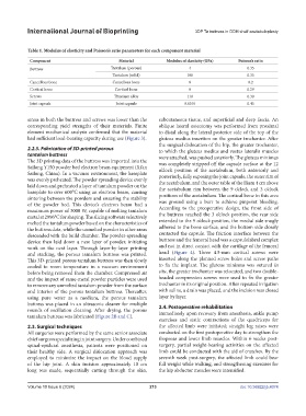
Table 1. Modulus of elasticity and Poisson’s ratio parameters for each component material
Component Material Modulus of elasticity (GPa) Poisson’s ratio
Buttress Tantalum (porous) 4 0.35
Tantalum (solid) 186 0.35
Cancellous bone Cancellous bone 8 0.2
Cortical bone Cortical bone 8 0.29
Screws Titanium alloy 110 0.30
Joint capsule Joint capsule 0.0105 0.45
stress in both the buttress and screws was lower than the subcutaneous tissue, and superficial and deep fascia. An
corresponding yield strengths of their materials. Finite oblique lateral osteotomy was performed from proximal
element mechanical analysis confirmed that the material to distal along the lateral posterior side of the top of the
had sufficient load-bearing capacity during use (Figure 3). gluteus medius insertion on the greater trochanter. After
the surgical dislocation of the hip, the greater trochanter,
2.2.5. Fabrication of 3D-printed porous to which the gluteus medius and vastus lateralis muscles
tantalum buttress were attached, was pushed anteriorly. The gluteus minimus
The 3D printing data of the buttress was imported into the was completely stripped off the capsule surface at the 12
Sailong Y150 powder bed electron beam equipment (Xi’an o’clock position of the acetabulum, both anteriorly and
Sailong, China). In a vacuum environment, the baseplate posteriorly, fully exposing the joint capsule, the outer rim of
was evenly preheated. The powder spreading device evenly the acetabulum, and the outer table of the ilium 4 cm above
laid down and preheated a layer of tantalum powder on the the acetabulum rim between the 9 o’clock and 3 o’clock
baseplate to over 600°C using an electron beam, causing
sintering between the powders and ensuring the stability positions of the acetabulum. The cortical bone in this area
of the powder bed. This device’s electron beam had a was ground using a burr to achieve pinpoint bleeding.
maximum power of 3000 W, capable of melting tantalum According to the preoperative design, the front side of
metal at 2996°C for shaping. The slicing software selectively the buttress reached the 3 o’clock position, the rear side
melted the tantalum powder based on the characteristics of extended to the 9 o’clock position, the medial side snugly
the buttress data, while the unmelted powder in other areas adhered to the bone surface, and the bottom side closely
descended with the build chamber. The powder-spreading contacted the capsule. The friction interface between the
device then laid down a new layer of powder, initiating buttress and the femoral head was a capsulolabral complex
work on the next layer. Through layer-by-layer printing and not in direct contact with the cartilage of the femoral
and stacking, the porous tantalum buttress was printed. head (Figure 4). Three 4.5-mm cortical screws were
This 3D-printed porous tantalum buttress was then slowly inserted along the planned screw holes and screw paths
cooled to room temperature in a vacuum environment to fix the implant. The gluteus minimus was sutured in
before being removed from the chamber. Compressed air situ, the greater trochanter was relocated, and two double-
and the impact of same-metal powder particles were used headed compression screws were used to fix the greater
to remove any unmelted tantalum powder from the surface trochanter in its original position. After repeated irrigation
and interior of the porous tantalum buttress. Thereafter, with saline, a drain was placed, and the incision was closed
using pure water as a medium, the porous tantalum layer by layer.
buttress was placed in an ultrasonic cleaner for multiple 2.4. Postoperative rehabilitation
rounds of oscillation cleaning. After drying, the porous
tantalum buttress was fabricated (Figure 2B and C). Immediately upon recovery from anesthesia, ankle pump
exercises and static contractions of the quadriceps for
2.3. Surgical techniques the affected limb were initiated; straight leg raises were
All surgeries were performed by the same senior associate conducted on the first postoperative day to strengthen the
chief surgeon specializing in joint surgery. Under combined iliopsoas and lower limb muscles. Within 6 weeks post-
spinal-epidural anesthesia, patients were positioned on surgery, partial weight-bearing activities on the affected
their healthy side. A surgical dislocation approach was limb could be conducted with the aid of crutches. By the
employed to minimize the impact on the blood supply seventh week post-surgery, the affected limb could bear
of the hip joint. A skin incision approximately 10 cm full weight while walking, and strengthening exercises for
long was made, sequentially cutting through the skin, the hip abductor muscles were intensified.
Volume 10 Issue 6 (2024) 213 doi: 10.36922/ijb.4074