
Page 237 - IJB-10-6
P. 237
International Journal of Bioprinting DIW of concave hydroxyapatite scaffolds
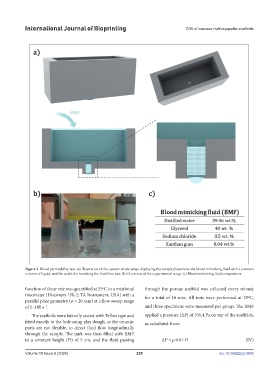
Figure 2. Blood permeability test. (a) Illustration of the custom-made setup, displaying the sample placement, the blood mimicking fluid with a constant
column of liquid, and the outlet for recording the fluid flow rate. (b) Overview of the experimental setup. (c) Blood mimicking fluid composition.
function of shear rate was quantified at 25ºC in a rotational through the porous scaffold was collected every minute
rheometer (Discovery HR-2; TA Instrument, USA) with a for a total of 10 min. All tests were performed at 25ºC,
parallel plate geometry (ø = 20 mm) at a flow sweep range
of 1–100 s . and three specimens were measured per group. The BMF
−1
The scaffolds were laterally sealed with Teflon tape and applied a pressure (∆P) of 331.4 Pa on top of the scaffolds,
fitted exactly to the hole using play dough, as the ceramic as calculated from:
parts are not flexible, to direct fluid flow longitudinally
through the sample. The tank was then filled with BMF
to a constant height (H) of 5 cm, and the fluid passing ∆P = ρ·9.81·H (IV)
Volume 10 Issue 6 (2024) 229 doi: 10.36922/ijb.3805