
Page 239 - IJB-10-6
P. 239
International Journal of Bioprinting DIW of concave hydroxyapatite scaffolds
samples for each type of geometry without cells were 3. Results and discussion
included, maintained over the culture period, stained, and 3.1. Scaffolds printability
decolorized. The supernatant of the control group was The three different types of TPMS (G, D, and S) designed
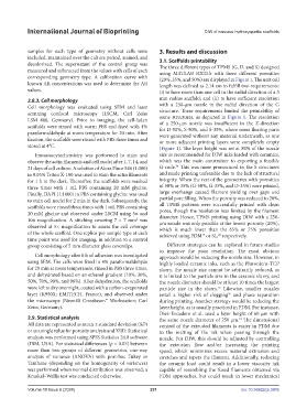
measured and subtracted from the values with cells of each using MATLAB R2021b with three different porosities
corresponding geometry type. A calibration curve with (20%, 35%, and 50%) are displayed in Figure 1. The unit cell
known AR concentrations was used to determine the AR length was defined as 2.14 cm to fulfill two requirements:
values. (i) to have more than one cell in the radial direction of a 3
2.8.3. Cell morphology mm radius scaffold; and (ii) to have sufficient resolution
Cell morphology was evaluated using SEM and laser with a 250-µm nozzle in the radial direction of the G
scanning confocal microscopy (LSCM; Carl Zeiss structure. These requirements limited the printability of
LSM 800, Germany). Prior to imaging, the cell-laden some structures, as depicted in Figure 1. The resolution
of a 250-µm nozzle was insufficient in the Z-direction
scaffolds were rinsed with warm PBS and fixed with 4% for D-50%, S-50%, and S-35%, where some floating parts
paraformaldehyde at room temperature for 20 min. After were generated without any material underneath, as one
fixation, the scaffolds were rinsed with PBS three times and or more adjacent printing layers were completely empty
stored at 4°C. (Figure 1). The layer height was set at 80% of the nozzle
Immunocytochemistry was performed to stain and size as recommended for DIW inks loaded with ceramics,
observe the actin filaments and cell nuclei after 1, 7, 14, and which was the main constrainer to exporting a feasible
54
21 days of cell culture. A solution of Alexa Fluor 546 (1:300) G-code. This was more pronounced in the S structures
in 0.05% Triton X-100 was used to stain the actin filaments and made printing unfeasible due to the lack of structural
for 1 h in the dark. Thereafter, the scaffolds were washed integrity. When the rest of the geometries with porosities
three times with 1 mL PBS containing 20 mM glycine. of 50% or 35% (G-50%, G-35%, and D-35%) were printed,
Finally, DAPI (1:1000) in PBS containing glycine was used large overhangs caused filament yielding over gaps and
to stain cell nuclei for 2 min in the dark. Subsequently, the partial pore filling. When the porosity was reduced to 20%,
scaffolds were rinsed three times with 1 mL PBS containing all TPMS patterns were successfully printed with clean
20 mM glycine and observed under LSCM using 5× and pores, though the resolution was limited by the filament
diameter. Hence, TPMS printing using DIW with a 250-
10× magnification. A stitching covering 7 × 7 mm was
2
observed at 5× magnification to assess the cell coverage µm nozzle was only possible at the lowest porosity (20%),
which is much lower than the 65% or 75% porosities
of the whole scaffold. One replica per sample type at each achieved using FDM or SL, respectively.
28
19
time point was used for imaging, in addition to a control
group consisting of 5 mm diameter glass coverslips. Different strategies can be explored in future studies
to improve the poor resolution. The most obvious
Cell morphology after 6 h of adhesion was investigated approach would be reducing the nozzle size. However, in
using SEM. The cells were fixed in 4% paraformaldehyde highly loaded ceramic inks, such as the Pluronic/α-TCP
for 20 min at room temperature, rinsed in PBS three times, slurry, the nozzle size cannot be arbitrarily reduced, as
and dehydrated based on an ethanol gradient (10%, 30%, it is linked to the particle size in the ceramic slurry, and
50%, 70%, 90%, and 96%). After dehydration, the scaffolds the nozzle diameter should be at least 10 times the largest
were left to dry overnight, coated with a carbon-evaporated particle size in the slurry. Likewise, smaller nozzles
55
layer (K950X; EMITECH, France), and observed under entail a higher risk of clogging and phase separation
56
the microscope (Neon40 Crossbeam™ Workstation; Carl during printing. Another strategy would be reducing the
Zeiss, Germany). layer height, as is usually practiced in FDM. For instance,
Diez-Escudero et al. used a layer height of 60 µm with
2.9. Statistical analysis the same nozzle diameter of 250 µm. The dimensional
19
All data are represented as mean ± standard deviation (SD) control of the extruded filaments is easier in FDM due
or as a single value for porosity analysis and XRD. Statistical to the melting of the ink when passing through the
analysis was performed using SPSS Statistics 24.0 software nozzle. For DIW, this should be adjusted by controlling
(IBM, USA). For statistical differences (p < 0.05) between the extrusion flow and/or increasing the printing
more than two groups of different geometries, one-way speed, which minimizes excess material extrusion and
analysis of variance (ANOVA) with post-hoc Tukey or stretches and tapers the filament. Additionally, reducing
Tamhane (depending on the homogeneity of variances) the ceramic load could result in a lower viscosity ink
was performed when normal distribution was observed; a capable of resembling the fused filaments obtained via
Kruskal–Wallis test was conducted otherwise. FDM approaches, but could result in lower mechanical
Volume 10 Issue 6 (2024) 231 doi: 10.36922/ijb.3805