Page 418 - IJB-10-6
P. 418
International Journal of Bioprinting                                 3D-bioprinted respiratory disease model


            collected nanoparticles were then freeze-dried for 24 h.   the material appeared homogeneous. HBEpCs were not
            To coat the nanoparticles, 0.1% chitosan (448877; Sigma-  added directly to the bioink and were instead seeded
            Aldrich, USA) was dissolved in 2% acetic acid and adjusted   onto the printed constructs seven days post-printing at a
            to pH 5 with NaOH (BP359, Lot 217634; Fisher Reagents,   density of 50,000 cells/construct.
            USA). Thereafter, 200 µL chitosan solution and 100 µL 5
            mM CaCl were added to the collected alginate particles,   2.4. Rheology
                    2
            mixed for 1 h, centrifuged, washed with ultrapure water   The rheological behavior of the cell-containing bioink was
            three times, and freeze-dried. As previously characterized,   tested and characterized using a TA Instruments HR20
            these nanoparticles were spherical with an average size of   Discovery Series Rheometer fitted with a 60 mm parallel
            just under 1000 nm. 33                             plate geometry. Constant shear stress sweeps (n = 3)
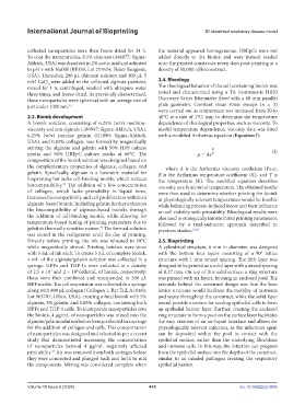
                                                               were carried out as temperature was increased from 20 to
            2.3. Bioink development                            40°C at a rate of 1°C/ min to determine the temperature
            A bioink solution, consisting of 6.25% (w/v) medium-  dependence of rheological properties, such as viscosity. To
            viscosity sodium alginate (180947; Sigma-Aldrich, USA),   model temperature dependence, viscosity data was fitted
            6.25% (w/v) porcine gelatin (G1890; Sigma-Aldrich,   with a modified Arrhenius equation (Equation I):
            USA), and 0.05% collagen, was formed by magnetically
            stirring  the  alginate and gelatin with  50% HPF  culture              B
            media and 50% HBEpC culture media at 60°C. The                    µ = Ae T                     (I)
            composition of the bioink solution was designed based on
            the complementary properties of alginate, collagen, and   where  A is the Arrhenius viscosity coefficient (Pa∙s),
            gelatin. Specifically, alginate is a favorable material for   B is the Arrhenius temperature coefficient (K), and T is
            bioprinting but lacks cell-binding motifs, which reduces   the temperature (K). The modified equation describes
            biocompatibility.   The  addition  of  a  low  concentration   viscosity as a function of temperature. The obtained results
                          45
            of collagen, which lacks printability in liquid form,   were then used to determine whether printing the bioink
            increases biocompatibility and cell proliferation within an   at physiologically relevant temperatures would be feasible
            alginate-based bioink. Including gelatin further enhances   while balancing process-induced forces and their influence
            the biocompatibility of alginate-based bioinks through   on cell viability with printability. Rheological results were
            the addition of cell-binding motifs, while allowing for   also used to strategically inform initial printing parameters,
            temperature-based tuning of printing parameters due to   followed by a trial-and-error approach described in
                                        45
            gelatin’s thermally sensitive nature.  The formed solution   previous studies. 33,45
            was stored in the refrigerator until the day of printing.
            Directly before printing, the ink was reheated to 50°C   2.5. Bioprinting
            while magnetically stirred. Printing batches were done   A cylindrical structure, 6 mm in diameter, was designed
            with 5 mL of ink each. To create 5 mL of complete bioink,   with the bottom four layers consisting of a 90° lattice
            4 mL of the alginate/gelatin solution was collected in a   structure with 1 mm strand spacing. The fifth layer was
            syringe.  HPFs and  THP-1s  were collected at  a density   designed to be printed as a solid layer with a strand spacing
            of 2.5 × 10  and 2 × 10 cells/mL of bioink, respectively;   of 0.27 mm. On top of this solid surface, a ring structure
                     7
                               6
            these were then combined and resuspended in 200 µL   was printed with six layers, forming an enclosed pool. The
            HPF media. The cell suspension was collected in a syringe   rationale behind the construct design was that the base
            along with 800 µL collagen (Collagen 1, Rat Tail, A10483,   lattice structure would facilitate the mobility of nutrients
            Lot 963787; Gibco, USA), creating a final bioink with 5%   and waste throughout the construct, while the solid layer
            alginate, 5% gelatin, and 0.05% collagen, containing both   would provide a surface for seeding epithelial cells to form
            HPFs and THP-1 cells. To incorporate nanoparticles into   an epithelial barrier layer. Further, creating the enclosed
            the bioink, 4 µg/mL of nanoparticles was mixed into the   ring structure to form a pool on the surface layer facilitates
            alginate/gelatin solution before being collected in a syringe   the easy creation of an air-liquid interface and allows for
            for the addition of collagen and cells. This concentration   physiologically relevant infection, as the infectious agent
            of nanoparticles was designed and selected as per a recent   can be deposited within the pool in contact with the
            study  that  demonstrated  increasing  the  concentration   epithelial surface, rather than the underlying fibroblasts
            of nanoparticles beyond 4 µg/mL negatively affected   and immune cells. In this way, the infection can progress
            printability.  Air was removed from both syringes before   from the epithelial surface into the depths of the construct,
                     33
            they were connected and plunged back and forth to mix   similar to an inhaled pathogen crossing the respiratory
            the components. Mixing was considered complete when   epithelial barrier.


            Volume 10 Issue 6 (2024)                       410                                doi: 10.36922/ijb.3895

