
Page 444 - IJB-10-6
P. 444
International Journal of Bioprinting Bioprinted skin scaffolds with GNP exposure
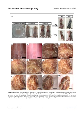
Figure 3. Transplantation on the dorsal skin of nude mice. (A) Illustration of the in vivo transplantation and administration processes. Layer-by-layer
structures were fabricated using the bioprinter and substituted the dorsal skin of the nude mice. One group was injected with GNPs and another group
was injected with saline. The entire bodies of mice from both groups were carefully monitored for 6 days before further experiments. (B–M) The recovery
process was illustrated by pictures of a mouse injected with saline with a fresh wound (B) and a scaffold transplanted into the wound directly after
implantation (C), as well as on days 1–10 (D–M). Scale bars: 200 μm. Abbreviations: GNP, gold nanoparticle.
Volume 10 Issue 6 (2024) 436 doi: 10.36922/ijb.4692