
Page 445 - IJB-10-6
P. 445
International Journal of Bioprinting Bioprinted skin scaffolds with GNP exposure
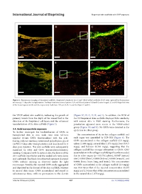
Figure 4. Fluorescence imaging of bioprinted scaffolds. Bioprinted constructs (A–C) and VEGF-added scaffolds (D–F) were captured by fluorescence
microscopy 11 days after transplantation. The bioprinted structures of pattern 2 (A and D) and pattern 3 (B and E) were imaged. (C and F) Magnified view
of the boxed regions in (B) and (E), respectively. Scale bars: 100 μm (A, B, D, and E); 20 μm (C and F).
the VEGF-added skin scaffolds, indicating the growth of (Figure 5C, D, G, H, K, and L). In addition, the ECM of
primary vessels from the depth of the wound bed in the the 3D-bioprinted skin scaffolds displayed little similarity
direction of the bioprinted cell layers and the enhanced with natural skin in H&E staining. Furthermore, the
vascularization of the skin scaffolds (Figure 5). penetration appeared more severe in the VEGF-added
group (Figure 5G and K). No GNPs were detected in the
3.5. Gold nanoparticle exposure epidermis in either group.
To further investigate the biodistribution of GNPs in
transplanted skin in vivo, nude mice were tail-vein The concentration of Au in the collagen scaffold and
injected 16-nm GNPs functionalized with the anti- each organ was quantified by ICP-OES (Figure 6). The
fouling polymer-methoxy-terminated polyethylene glycol GNPs accumulated in the collagen scaffold (2.5 mg/g),
(mPEG) 5 days after transplantation and monitored for 6 spleen (1.606 mg/g), natural skin (1.070 mg/g), liver (0.915
days post-injection. The skin scaffolds were subsequently mg/g), and kidneys (0.364 mg/g), suggesting that the
examined by H&E and CD31 immunohistochemistry collagen scaffold has stronger adsorption to GNPs. GNP
staining. Compared with the natural skin, the dermis of the accumulation in the collagen scaffold significantly exceeded
skin scaffolds was thicker and the seeded cells were dense that in other organs (p = 0.0174 [spleen], 0.0047 [natural
and uniformly. The black dots observed represent clustered skin], 0.0042 [liver], 0.0008 [kidney], 0.0005 [muscle], and
GNPs without staining as observed under the light 0.0004 [bone, heart, lung, and brain]). The concentration
microscope. Notably, the injected GNPs easily aggregated of GNPs accumulated in the collagen scaffold (2.5 mg/g)
and deposited in the dermis of the skin scaffolds, whereas was 10.6 times that of the injected concentration (0.235
in natural skin tissue, GNPs accumulated and stayed in mg/g) and 2.3 times that of the concentration accumulated
subcutaneous tissue with no permeation to the dermis in the natural skin (1.070 mg/g).
Volume 10 Issue 6 (2024) 437 doi: 10.36922/ijb.4692