
Page 520 - IJB-10-6
P. 520
International Journal of Bioprinting Redefined collagen inks in cartilage printing
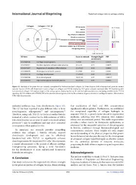
Figure 13. Analysis of the genes that are uniquely upregulated by biofunctionalized collagen. (a) Venn diagram (left) of upregulated genes in stromal
vascular fraction (SVF) cells bioprinted in pure collagen or collagen and TGF-β, displaying 1057 genes uniquely expressed after 3D bioprinting with
functionalized collagen. GO analysis (right) of the unique genes, displaying the top 10 enriched pathways/processes, including enrichment for TGF-β
signaling. (b) GO analysis with STRING DB of the abovementioned genes confirms the activation of genes involved in cartilage formation. Abbreviation:
FDR: False discovery rate.
undesired pathways (e.g., bone development; Figure 13). that modulation of NaCl and PBS concentrations
This GF has been reported to play different roles in bone significantly affects gelation. Furthermore, we established
morphogenesis, adipogenesis, and carcinogenesis. the exceptional capability of collagen hydrogels to
56
55
Therefore, using only TGF-β1 to biofunctionalize bioinks sequester TGF-β1, a growth factor pivotal in regenerative
instead of a whole cocktail for the differentiation of MSCs medicine, achieving over 99% retention with minimal
into chondrocytes (as commonly used in standard culture release over an extended period. This stable sequestration
protocols ) may be insufficient and may elicit unwanted provides a robust matrix for therapeutic applications, as
57
consequences when applied in vivo. evidenced by the successful induction of chondrogenesis
in bioinks, confirmed through histomorphological and
In summary, our research provides compelling transcriptomic analyses. These insights not only deepen
evidence that collagen I bioinks robustly support our understanding of the physical properties that govern
chondrocyte development and can be effectively bioink behavior but also pave the way for the development
functionalized with TGF-β1 to significantly enhance of advanced collagen-based hydrogels as effective carriers
chondrogenesis in 3D-bioprinted SVF cells. This represents for the controlled delivery of bioactive molecules,
a crucial advancement in the pursuit of efficient cartilage progressing the field of tissue engineering and regenerative
bioengineering processes, laying a solid foundation medicine.
for the development of more sophisticated biomaterial
formulation strategies. Acknowledgments
4. Conclusion The authors would like to thank (i) Prof. S. Todinova from
the Institute of Biophysics and Biomedical Engineering,
Our study underscores the neglected role of ionic strength Bulgarian Academy of Sciences, for her assistance with DSC
in the gelation kinetics of collagen bioinks, demonstrating analysis, and (ii) Assoc. Prof. L. Simova from the Institute
Volume 10 Issue 6 (2024) 512 doi: 10.36922/ijb.4566