
Page 516 - IJB-10-6
P. 516
International Journal of Bioprinting Redefined collagen inks in cartilage printing
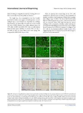
demonstrating an insignificant number of dying cells even Next, we assessed the morphology of the SVF cells
three weeks after bioprinting (data not shown). compared to chondrocytes in terms of the formation of
lacunae, secretion of proteoglycans (Alcian blue staining),
The study was then expanded to test the bioinks’ and collagen type II (IHC). We found that both bioinks
potential to induce and support chondrogenesis, as both induced chondrogenesis in at least a portion of the SVF
collagen type I and TGF-β1 are important for cartilage cells as observed from H&E and staining for typical
development. To assess this, we used an in vitro model cartilage ECM components. Notably, weaker staining for
cell line of MSCs derived from white adipose tissue (or proteoglycans and collagen II was observed in the collagen
SVF cells), hTERT A41hWAT-SVF. Cells in both bioinks bioink compared to CHON-001 (printed in collagen) and
demonstrated good viability of around 90% on day 1 SVF cells in the biofunctionalized bioink (Figure 10b),
and 80% on day 14, even if there were more dying cells suggesting that TGF-β1 release enhances the chondrogenic
compared to CHON-001 (Figure 10a). properties of the biomaterial.
Figure 10. Evaluation of chondrogenesis support of pure and biofunctionalized collagen bioinks. (a) Live-dead staining of CHON-001 cells (left) and
stromal vascular fraction (SVF) cells in pure collagen (middle) and in collagen biofunctionalized with TGF-β (right) on days 1 (top panel) and 14 (bottom
panel). (b) Histomorphology of chondrogenesis in CHON-001 (left column) and SVF cells in pure collagen (middle column) and biofunctionalized
collagen (right column) through hematoxylin and eosin (H&E) staining, staining with Alcian blue for proteoglycans and glycosaminoglycans, and
immunohistochemistry (IHC) for the cartilage biomarker collagen II (Col II) (top, middle, and bottom rows, respectively). The inset images denote regions
of interest that are at 2× higher magnification (digital for (a) and taken with a 20× objective for (b).
Volume 10 Issue 6 (2024) 508 doi: 10.36922/ijb.4566