
Page 514 - IJB-10-6
P. 514
International Journal of Bioprinting Redefined collagen inks in cartilage printing
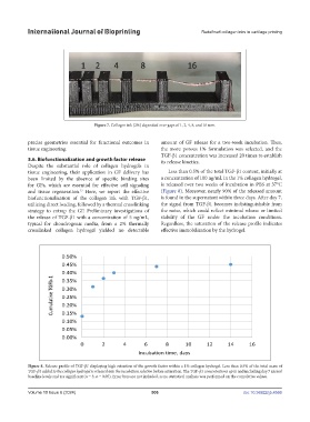
Figure 7. Collagen ink (2%) deposited over gaps of 1, 2, 4, 8, and 16 mm.
precise geometries essential for functional outcomes in amount of GF release for a two-week incubation. Thus,
tissue engineering. the more porous 1% formulation was selected, and the
TGF-β1 concentration was increased 20 times to establish
3.6. Biofunctionalization and growth factor release its release kinetics.
Despite the substantial role of collagen hydrogels in
tissue engineering, their application in GF delivery has Less than 0.5% of the total TGF-β1 content, initially at
been limited by the absence of specific binding sites a concentration of 100 ng/mL in the 1% collagen hydrogel,
for GFs, which are essential for effective cell signaling is released over two weeks of incubation in PBS at 37°C
and tissue regeneration. Here, we report the effective (Figure 8). Moreover, nearly 90% of the released amount
27
biofunctionalization of the collagen ink with TGF-β1, is found in the supernatant within three days. After day 7,
utilizing direct loading, followed by a thermal crosslinking the signal from TGF-β1 becomes indistinguishable from
strategy to entrap the GF. Preliminary investigations of the noise, which could reflect minimal release or limited
the release of TGF-β1 with a concentration of 5 ng/mL, stability of the GF under the incubation conditions.
typical for chondrogenic media, from a 2% thermally Regardless, the saturation of the release profile indicates
crosslinked collagen hydrogel yielded no detectable effective immobilization by the hydrogel.
Figure 8. Release profile of TGF-β1 displaying high retention of the growth factor within a 1% collagen hydrogel. Less than 0.5% of the total mass of
TGF-β1 added to the collagen hydrogel is released into the incubation solution before saturation. The TGF-β1 concentrations up to and including day 7 exceed
baseline levels and are significant (n = 3; α = 0.05). Error bars are not included, as no statistical analysis was performed on the cumulative values.
Volume 10 Issue 6 (2024) 506 doi: 10.36922/ijb.4566