
Page 15 - IJB-7-1
P. 15
Zhang, et al.
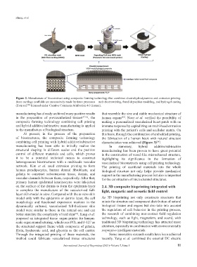
Figure 2. Manufacture of bioconstruct using composite forming technology that combines electrohydrodynamics and extrusion printing.
Bone cartilage scaffolds are successively made by three processes – melt electrowriting, fused deposition modeling, and hydrogel casting
(from ref. licensed under Creative Commons Attribution 4.0 license).
[45]
manufacturing has already archived many positive results that resemble the size and stable mechanical structure of
in the preparation of prevascularized tissues [57-61] , the human organs . Noor et al. verified the possibility of
[63]
composite forming technology combining cell printing making a personalized vascularized heart patch with no
and hybrid additive/subtractive manufacturing is applied immune response by capitalizing on multi-head extrusion
to the manufacture of biological structure. printing with the patient’s cells and acellular matrix. On
At present, in the process of the preparation this basis, through the combination of embedded printing,
of biostructures, the composite forming technology the fabrication of a human heart with natural structure
combining cell printing with hybrid additive/subtractive characteristics was achieved (Figure 3) .
[64]
manufacturing has been able to initially realize the In summary, hybrid additive/subtractive
structural shaping in different scales and the position manufacturing has been proven to have great potential
control of different materials and cells, which proves in the construction of vessel-like microchannel structure,
it to be a potential technical means to construct highlighting its significance in the formation of
heterogeneous biostructures with a multiscale vascular vascularized bioconstructs using cell printing technology.
network. Kim et al. used extrusion printing to form The printing of sacrificial materials into the whole
human preadipocytes, human dermal fibroblasts, and biological structure not only helps provide mechanical
gelatin to construct subcutaneous tissue, dermis, and support in the manufacturing process but also is important
vascular channels between them, respectively. After that, for the construction of microchannel structures.
primary human epidermal keratinocytes were ink-jetted
on the surface of the dermis to form the epidermis layer 2.4. 3D composite bioprinting integrated with
to complete the manufacture of the vascularized full- light, magnetic and acoustic field control
layer skin model in vitro. Compared with the in vitro skin
model only with the epidermis or dermis layer, the cell As 3D bioprinting not only constructs structures that
morphology and functional expression markers in the mimic the structure and component distribution of natural
dynamically cultured, vascularized full-thickness skin biological tissues and organs but also take into account
model were similar to those in the natural one and can the regulation of cell behavior in the printing process,
better simulate the complexity of real skin . Kang et al. the research of combining non-contact field regulation
[62]
proposed an integrated tissue–organ printer for human- technology, such as light, magnetism, and sound, with
scale organ manufacturing, which uses PCL and F-127 as traditional 3D bioprinting technology has attracted more
the structural support frame while composite of gelatin, attention, especially in combination with environmentally
fibrin, hyaluronic acid, and glycerin as the cell carrier. responsive intelligent materials.
Through the integrated printing of these materials, the Some innovative research results have been achieved
method could fabricate vascularized tissue structures recently. Yang et al. combined the external DC electric
International Journal of Bioprinting (2021)–Volume 7, Issue 1 11