
Page 24 - IJB-7-2
P. 24
Additively Manufactured NiTi Implants
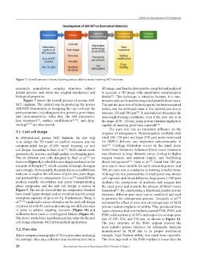
Figure 7. Overall process of manufacturing porous additive manufacturing-NiTi implants.
accurately manufacture complex structures without 2D image, and then the data must be compiled and analyzed
further process and retain the original mechanical and to generate a 3D image with quantitative morphological
biological properties. details . This technique is attractive because it is non-
[76]
Figure 7 shows the overall process of porous AM- invasive and can be used to image and quantify bone repair.
NiTi implants. The critical step in producing the porous The optimal pore size of hydroxyapatite has been measured
AM-NiTi biomaterials is designing the unit cell and the before, and the estimated value of the optimal pore size is
pore parameters, including pore size, porosity, pore shape, between 150 and 500 μm . It was believed that under the
[77]
and interconnectivity. After that, the AM parameters, non-weight-bearing conditions, even if the pore size is in
heat treatment , surface modification [66-68] , and drug- the range of 50–125 μm, using porous titanium implants is
[15]
loading [69,70] are also needed. capable of ensuring good bone ingrowth .
[78]
The pore size has an important influence on the
3.1. Unit cell design progress of osteogenesis. Hydroxyapatite scaffolds with
In AM-produced porous NiTi implants, the first step small (90–120 μm) and large (350 μm) pores were used
is to design the 3D model of scaffold structure and the for BMP-2 delivery and implanted subcutaneously in
[50]
computer-aided design (CAD) model focusing on unit rats . Cartilage formation occurs in the small pores
cell design. According to Bael et al. , SLM cannot create before bone formation. Enhanced blood vessel formation
[71]
geometrically accurate and high-quality overhanging parts. was observed in large diameter pores, leading to higher
The six different unit cells designed by Bael et al. are oxygen tension and nutrient supply, and facilitating
[72]
shown in Figure 8, in which the pore shapes are based on the direct osteogenesis . Gotz et al. found that 200 μm
[50]
[79]
research of Rumpler , which consists of triangle hexagon pore size is more suitable for early osseointegration, and
[73]
and rectangle. Subsequently, the printed porous scaffold was 300 μm pore size is conducive to forming lamellar bone.
analyzed to explore the influence of pore size, pore shape, Although the low permeability of small pores may hinder
and permeability on osteogenesis. Li et al. used SLM to cell ingrowth and blood diffusion, large pores (>300 μm)
[74]
produce metallic microlattice and epoxy interpenetrating facilitate the absorptions of nutrients and oxygen into
phase composites and the unit cell design is shown in the inner pores and promote the process of blood vessel
Figure 9. The results showed that the composites obtained formation . By constructing a functional graded porous
[65]
have much higher strength and an excellent specific energy structure, different pore sizes can be combined and used
absorption capacity of up to 46 J/g. Furthermore, Wang et to promote the osteogenesis process. Taniguchi et al.
[80]
al. [22,75] conducted a series of studies on the unit cell design evaluated the effect of pore size on osteogenesis of SLM
of porous Ti-6Al-4V, such as introducing two different sizes porous titanium implants in rabbits. They produced three
of pores to achieve coupling functions (Figure 10) and types of porous titanium implants named P300, P600, and
millimeter-level pores in novel gyroid lattices (Figure 11). P900, with a porosity of 65% and respective average pore
The above works have significant guiding value for the unit size of 309, 632, and 956 μm, as shown in Figure 12.
cell design of porous NiTi in biomedical applications. The pore structure of the P600 implant showed the
3.2. Pore size most suitable porous structure for orthopedic implants
manufactured by SLM due to its proper mechanical
Micro-computed tomography (CT) is a procedure including strength, high fixation ability, and rapid bone ingrowth.
the isotropic slice data collection and reconstruction into a The bone ingrowth of the P300 implant is lower than the
20 International Journal of Bioprinting (2021)–Volume 7, Issue 2