
Page 25 - IJB-7-2
P. 25
Zhang, et al.
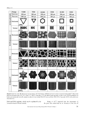
Figure 8. (A) Unit cells. (B) Optical microscopy images. (C) SEM images. (D) Models based on micro-computed tomography (Reprinted
[72]
from Acta Biomaterialia, 8(7), B. S. Van, Y. C. Chai, S. Truscello, et al., the effect of pore geometry on the in vitro biological behavior of
human periosteum-derived cells seeded on selective laser-melted Ti6Al4V bone scaffolds, 2824–2834, Copyright (2012), with permission
from Elsevier).
P600 and P900 implants, which can be explained by the Wang et al. reported that the increment of
[81]
formation level of blood vessels. the pore size could lead to an increase in the size of
International Journal of Bioprinting (2021)–Volume 7, Issue 2 21