
Page 26 - IJB-7-2
P. 26
Additively Manufactured NiTi Implants
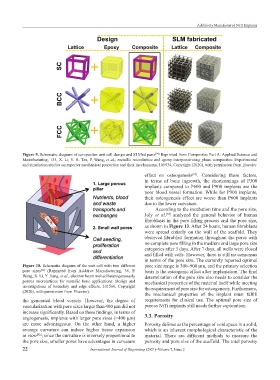
Figure 9. Schematic diagram of composites unit cell design and SLMed parts Reprinted from Composites Part A: Applied Science and
[74]
Manufacturing, 135, X. Li, Y. H. Tan, P. Wang, et al., metallic microlattice and epoxy interpenetrating phase composites: Experimental
and simulation studies on superior mechanical properties and their mechanisms, 105934, Copyright (2020), with permission from Elsevier.
effect on osteogenesis . Considering these factors,
[83]
in terms of bone ingrowth, the shortcomings of P300
implants compared to P600 and P900 implants are the
poor blood vessel formation. While for P900 implants,
their osteogenesis effect are worse than P600 implants
due to the lower curvature.
According to the incubation time and the pore size,
Joly et al. analyzed the general behavior of human
[84]
fibroblasts in the pore filling process and the pore size,
as shown in Figure 13. After 24 hours, human fibroblasts
were spread entirely on the wall of the scaffold. They
observed fibroblast formation throughout the pores with
no complete pore filling in the medium and large pore size
categories after 3 days. After 7 days, all wells were closed
and filled with cells. However, there is still no consensus
in terms of the pore size. The currently reported optimal
Figure 10. Schematic diagram of the unit cell with two different pore size range is 300–900 μm, and the primary selection
pore sizes (Reprinted from Additive Manufacturing, 36, P. basis is the osteogenic effect after implantation. The final
[22]
Wang, X. Li, Y. Jiang, et al., electron beam melted heterogeneously determination of the pore size also needs to consider the
porous microlattices for metallic bone applications: Design and mechanical properties of the material itself while meeting
investigations of boundary and edge effects, 101566, Copyright
(2020), with permission from Elsevier). the requirement of pore size for osteogenesis. Furthermore,
the mechanical properties of the implant must fulfill
the generated blood vessels. However, the degree of requirements for clinical use. The optimal pore size of
vascularization with pore sizes larger than 400 μm did not porous NiTi implants still needs further exploration.
increase significantly. Based on these findings, in terms of
angiogenesis, implants with larger pore sizes (>400 μm) 3.3. Porosity
are more advantageous. On the other hand, a higher Porosity defines as the percentage of void space in a solid,
average curvature can induce higher tissue expansion which is an inherent morphological characteristic of the
in vitro ; since the curvature is inversely proportional to material. There are different methods to measure the
[82]
the pore size, smaller pores have advantages in curvature porosity and pore size of the scaffold. The total porosity
22 International Journal of Bioprinting (2021)–Volume 7, Issue 2