
Page 28 - IJB-7-2
P. 28
Additively Manufactured NiTi Implants
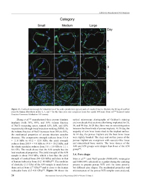
Figure 13. Confocal microscopy for visualization of the actin cytoskeleton (green) and cell nuclei (blue) to illustrate the filling of scaffold
pores by human fibroblasts at day 1, 3, and 7 for the three pore size categories (scale bar equals 100 mm) (from ref. licensed under
[84]
Creative Commons Attribution 3.0 license).
Zheng et al. manufactured three porous titanium optical microscope photographs of Goldner’s staining
[88]
implants (with 30%, 40%, and 50% volume fraction and non-decalcified sections after being implanted for 28,
of NaCl occupying space, named A30, A40, and A50, 56, and 84 day. At 28 day, there was no osseointegration
respectively) through metal injection molding (MIM). As between the three kinds of porous implants. At 56 day, the
the volume fraction of NaCl increases from 30% to 50%, majority of new bone tissue stuck to the implant surface.
the mechanical properties of porous titanium samples At 84 day, the porous implant and the host bone tissue
decrease. The compressive strength reduces from 316.6 were tightly bonded. The deep and surface pores of the
± 11.4 MPa to 63.2 ± 12.8 MPa, the yield strength porous implant are overgrown with vascular-like tissue
reduces from 284.9 ± 9.8 MPa to 59.8 ± 10.2 MPa, and and mineralized bone matrix. The bone tissues of the
the elastic modulus reduces from 3.0 ± 1.0 GPa to 1.1 ± A40 and A50 groups were deeper than those of the A30
[88]
0.6 GPa. The result shows that the A30 sample has the group .
best mechanical properties. The yield strength of the A50
sample is about 59.8 MPa, which is less than the yield 3.4. Pore shape
strength of cortical bone (80–120 MPa) and close to that Irfan et al. used NaCl powder (N400-60% rectangular
[91]
of human trabecular bone (0.2–80 MPa) . The modulus and N400-60% spherical) as a gasket during the sintering
[89]
of elasticity (1.1 GPa) of the A50 sample is much lower process to prepare porous NiTi with the same porosity
than cortical bone (17 GPa) and is close to the human but different pore shapes. The mechanical properties and
[90]
trabecular bone (0.5–4.0 GPa) . Figure 14 shows the microstructure of the porous NiTi samples were analyzed,
[89]
24 International Journal of Bioprinting (2021)–Volume 7, Issue 2