
Page 64 - IJB-7-2
P. 64
3D Bioprinted Triple-layered Human Alveolar Lung Models
laden droplets using DOD bioprinting technique enables A549 epithelial cells cultured under submerged condition
the cells to proliferate and spread uniformly to form exhibited clear cell edges and organelles and covered more
a homogeneous cell layer at day 7. As the printed cells than 70% surface of the 3D alveolar lung tissue models
were close to 90% confluence by day 7 for both A549 and after 3 days of culture under LLI condition. Conversely,
EA.hy926, further proliferation studies for all 3 types of the A549 epithelial cells cultured under ALI condition
cells were not continued. As such, we have demonstrated started to flatten and formed compacted layer initially
the use of microvalve-based bioprinting technique can before forming some spheroid-like structures on top of the
achieve consistent printed cell output with high short- flattened cell layer over time. The observations from our
term (>97%) and long-term viability (over a period of work are corroborated by other studies that characterized
at least 7 days) using the PVP-modified cell suspension. the mono-culture of A549 cells at ALI interface [72,73] .
This is critical for 3D DOD bioprinting of different human In general, the 3D bioprinted alveolar lung models
alveolar lung cells to achieve precise and uniform cell show high viability (>96%) over a period of 14 days. It
deposition and patterning within the 3D tissue constructs is noted that the overall viability of the 3D bioprinted
to achieve high repeatability at high-throughput rates. human alveolar lung models is higher at day 10 and 14 as
compared to day 7; this is likely due to cell proliferation
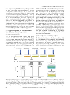
3.3. Characterization of 3D bioprinted triple- over time that led to an increase in the total number
layered human alveolar lung models of cells, resulting in a lower ratio of dead cells to total
number of cells (Figure 4B).
(A) Long-term survivability
The 3D triple-layered human alveolar lung model (2) Immunofluorescence staining analysis
consisted of A549 human lung epithelial cells (top), To determine the influence of culture conditions (ALI
EA.hy926 human endothelial cells (middle), and MRC-5 vs. LLI conditions) on the maturation of 3D bioprinted
human lung fibroblasts (bottom). The sequence of printing human alveolar lung models, the lung tissue models
was as follows: Collagen >MRC-5 >EA.hy926 >Collagen were stained for the presence of alveolar type I (AT-1)
>A549 (CMECA) to mimic the spatial arrangement of biomarkers (aquaporin-5, caveolin-1), alveolar type II
native lung alveolar cells and its ECM (Figure 4A). The (AT-2) biomarker (Pro-SPC), epithelial biomarker (pan-
A
B
Figure 4. (A) Bioprinting process of the 3D triple-layered human alveolar lung models; the ECM bio-inks and cell droplets are deposited
in the following order: Collagen >MRC-5 >EA.hy926 >Collagen >A549 to mimic the spatial arrangement of native alveolar blood-air
lung cells from the basal to apical layers, and its ECM. The 3D bioprinted blood-air barrier models are then cultivated under liquid-liquid
interface (submerged condition) for the first 3 days followed by air-liquid interface up to additional 11 days. (B) The graph shows the
survivability results of the triple-layered blood-air barrier models (CMECA) over a culture period of 14 days based on NucFix staining.
60 International Journal of Bioprinting (2021)–Volume 7, Issue 2