
Page 61 - IJB-7-2
P. 61
Ng, et al.
A D
B
C E
F
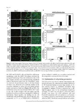
Figure 1. (Left) Representative fluorescence images of (A) A549 human lung epithelial cells, (B) EA.hy926 human endothelial cells, and
(C) MRC-5 human lung fibroblasts (bottom), cultured in different culture medium (co-culture medium and control medium – A549 with
RPMI-1640 culture medium supplemented with 10% fetal bovine serum [FBS] and both EA.hy926 and MRC-5 with DMEM/F12 culture
medium supplemented with 10% FBS) over a period of 7 days – the green fluorescent represents the viable cells while the blue fluorescent
represent the cell nuclei; scale bar: 100 µm. (Right) Influence of co-culture medium on proliferation profile of (D) A549 human lung
epithelial cells, (E) EA.hy926 human endothelial cells, and (F) MRC-5 human lung fibroblasts over a period of 7 days.
the A549 and EA.hy926 cells exhibited the cobblestone culture medium is suitable as a co-culture medium and
morphology, while the MRC5 fibroblasts exhibited the this composition was used for the whole study.
elongated morphology. Next, the proliferation rates of all
three human cell lines were measured using normalized 3.2. Optimization of cell printing parameters
relative fluorescence units from the PrestoBlue assay. The next critical step was to evaluate and determine the
®
As observed, the proliferation rates of all three human printed cell output for consistent and scalable fabrication
cell lines were observed to be relatively similar in the of 3D bioprinted human alveolar lung models. A 30-
co-culture (indicated as white bar) and original growth min printing window is considered reasonably long
media (indicated as black bar) (Figure 1B and 1C). The for printing of multiple 3D tissue constructs due to the
proliferation assay was not evaluated further after 7 days high-throughput rates for the jetting-based bioprinting
as the cells would overgrow and start aging, and this technique. To mimic the cell density within the native
would not be able to provide any meaningful or additional alveolar lung tissue, the A549 epithelial cells and
information. Taken together, our findings on the cell EA.hy926 endothelial cells were printed at a cell density
morphology and proliferation rate showed that the ratio of 2 million cells/ml to create densely-packed cell layers
composition of 1:1 v/v of RPMI-1640 to DMEM/F12 while the MRC5 fibroblasts were printed at a cell density
International Journal of Bioprinting (2021)–Volume 7, Issue 2 57