
Page 15 - IJB-7-3
P. 15
Liu, et al.
A C
B D
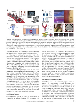
Figure 4. Devices developed for bioprinting microvessels. (A) Microvascular structures could be built by bioprinting based on inkjet,
extraction, and direct laser writing (from ref. licensed under Creative Commons Attribution License. Copyright © Mary Ann Liebert).
[77]
(B) Direct bioprinting of microvascular structures based on the coaxial nozzle, and microscopic view of L929 mouse fibroblasts encapsulated
by tubular alginate (Republished with permission from reference ). (C) Experimental setup and fabrication of engineered tissues containing
[88]
[93]
microvascular structures using optical stereolithography . (D) Microscopic photograph of a microheater array used to perform thermal
stereolithography (Republished with permission, from Kojima M, Horade M, Takata S, et al., IEEE International Conference on Cyborg
and Bionic Systems, IEEE, 2018. ).
[97]
bioprinting. However, its disadvantages are low cell density built-in microchannels by controlling the crosslinking
and low mechanical properties of the printed structures. time sequence and using the fusion of adjacent hollow
Targeting to printing the materials with better filaments. As shown in Figure 4B, a novel 3D bioprinting
mechanical properties, researchers developed extrusion method was proposed for fabricating cell-loaded built-
bioprinting to deposit viscous materials . The extrusion in hollow hydrogel structures in the study by Arai et al.
[82]
is commonly actuated by air pump screw plunger. Similar and Gao et al. [88,89] . The inner diameter of the fabricated
to inkjet bioprinting, extrusion bioprinting is also capable microvessels could be smaller than 200 μm and longer
of composite bioprinting with multiple bioinks. It also than 10 cm. Coaxial nozzles are used to fabricate hollow
allows high cell density. However, the relatively low cell alginate fibers that are able to move in the XY direction,
viability limited its application, since the large pressure is with a Z-shaped platform, and raw materials such as
harmful to the living cells [83-85] . calcium chloride solution to precisely control the XY
Laser-assisted bioprinting is based on laser direct direction to determine the coaxial nozzle position for
writing and laser-induced transfer techniques [86,87] . The planar feature printing. In contrast to other bioprinting
laser-assisted bioprinting devices’ core is a three-layer methods used to fabricate built-in microchannel
laser-responsive plate. The plate contains a top energy- structures, this method allows for simultaneous printing
absorbing layer, a middle donor layer, and a bottom bioink of scaffolds and microchannels [88-90] .
layer. When the focused laser is at a small local area of
the energy-absorbing layer, a small part of the donor layer 5.3. Optical stereolithography
under the laser exposure will be vaporized to form a high- Bioprinting based on stereolithography technique
pressure microbubble. The bubble will impel the bioinks, belongs to layer-by-layer printing featuring high
and the formed small droplet of bioink will fall onto the efficiency. Stereolithography is a technique applying
substrate. In laser-assisted bioprinting, cells are protected the selective solidification of curable bioinks [91,92] . As
from the damages of the high pressure, thereby achieving shown in Figure 4C, the digital mirror device (DMD) is
high cell viability. However, the system is complex and most commonly utilized in optical stereolithography to
expensive. Its unable to print multiple materials. pattern the 2D parallel light. The printing resolution can
5.2. Coaxial nozzle be as high as 1 μm. Compared with the extrusion-based
bioprinting using high pressure, optical stereolithography
Considering the special structure of microvessels, it can achieve relatively higher cell viability. In the process
is possible to prepare hydrogel 3D structures with of engineering the microvessels using this method,
International Journal of Bioprinting (2021)–Volume 7, Issue 3 11