
Page 62 - IJB-7-3
P. 62
Heart-on-a-chip
A B
C D
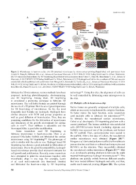
Figure 2. Microtissues in heart-on-a-chip. (A) 2D patterned microtissues by micro-contact printing (Republished with permission from
Annabi N, Tsang K, Mithieux SM, et al., Advanced Functional Materials, © 2013 WILEY‐VCH Verlag GmbH and Co. KGaA, Weinheim).
(B) 3D vascularized microtissue by 3D bioprinting (Republished with permission from Colosi C, Shin SR, Manoharan V, et al., Advanced
Materials, © 2015 WILEY‐VCH Verlag GmbH and Co. KGaA, Weinheim.) (C) 3D hydrogel scaffold for the co-culture of CMs and vascular
endothelial cells (Republished with permission from Morgan KY, Sklaviadis D, Tochka ZL, et al., Advanced Functional Materials, © 2016
WILEY‐VCH Verlag GmbH and Co. KGaA, Weinheim.) (D) Microtissues with multiple cells by 3D bioprinting (Republished with permission
from Miri AK, Daniel N, Luis I, et al., Adv Mater, ©2018 WILEY‐VCH Verlag GmbH and Co. KGaA, Weinheim)
fabricate the 3D microtissues, various methods have been technology . Using this chip, the alignment of cells can
[12]
proposed, including photolithography, electrospinning, be well controlled by fabricating some microgrooves in
and 3D bioprinting. Among them, 3D bioprinting the chip.
is considered a promising technique to fabricate 3D
microtissues. The cell-laden bioinks are printed through (3) Multiple cells in heart-on-a-chip
the nozzle of a 3D printer. Bioinks are of great importance Native tissues are generally composed of multiple cells,
for 3D bioprinting of microtissues. So far, the most which are necessary to implement the complex functions.
frequently used bioinks are hydrogels. Hydrogels have To better mimic the heart function, some researchers
similar structure with ECM and porous properties as used multiple cells to fabricate the microtissues [29,30] .
well as good diffusion of biomolecules. Thus, they are To fabricate the vascularized cardiac microtissues,
promising candidates for the fabrication of microtissues Colosi et al. developed a 3D bioprinting platform with a
and simulation of the growth environment for cardiac coaxial printhead. The human umbilical vein endothelial
cells in vivo . Their stiffness is adjustable and the
[27]
biocompatibility is good after modification. cells (HUVECs)-laden bioink (including alginate and
Some researchers used 3D bioprinting to GelMA) was squeezed out of the printhead, and formed
fabricate microtissues in heart-on-a-chip. Chen et al. the 3D scaffold. Then, cardiomyocytes were seeded in
prepared CMs-laden GelMA and fabricated the cardiac the scaffold. After a few days of culture, the vascularized
[31]
microtissues using 3D bioprinting . The phonotype of cardiac microtissues were obtained (Figure 2B) .
[28]
cells is similar with the cells in vivo. Hydrogel based 3D Morgan et al. fabricated polymer scaffold with multiscale
bioprinting has shown a great potential in fabrication of porous structure and then co-cultured cardiomyocytes and
microtissues. Due to the good biocompatibility, hydrogel- HUVECs on the structure. They successfully obtained
[32]
based microtissues provide ideal microenvironments for the vascularized cardiac microtissue (Figure 2C) . Miri
cell growth and proliferation. Recently, some researchers et al. developed a printing platform to fabricate the multi
[33]
have attempted to print the microtissues together with material heterogeneous hydrogel microstructures . This
microfluidic chips in one step. For example, Lewis platform can quickly switch between different nozzles
et al. used multi-materials (six functional bioinks) that have loaded different hydrogels and cells, and thus,
and fabricated the heart-on-a-chip by 3D bioprinting enable multi-material printing (Figure 2D). The system
58 International Journal of Bioprinting (2021)–Volume 7, Issue 3