
Page 11 - IJB-8-2
P. 11
Hu, et al.
A B C
D E F
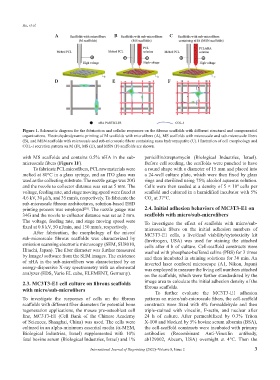
Figure 1. Schematic diagram for the fabrication and cellular responses on the fibrous scaffolds with different structural and componential
organizations. Electrohydrodynamic printing of M scaffolds with microfibers (A), MS scaffolds with microscale and sub-microscale fibers
(B), and MSN scaffolds with microscale and sub-microscale fibers containing nano hydroxyapatite (C). Illustration of cell morphology and
COL-I secretion pattern on M (D), MS (E), and MSN (F) scaffolds are shown.
with MS scaffolds and contains 0.5% nHA in the sub- penicillin/streptomycin (Biological Industries, Israel).
microscale fibers (Figure 1F). Before cell seeding, the scaffolds were punched to have
To fabricate PCL microfibers, PCL raw materials were a round shape with a diameter of 15 mm and placed into
melted at 80°C in a glass syringe, and an ITO glass was a 24-well culture plate, which were then fixed by glass
used as the collecting substrate. The nozzle gauge was 20G rings and sterilized using 75% alcohol aqueous solution.
and the nozzle to collector distance was set as 5 mm. The Cells were then seeded at a density of 5 × 10 cells per
4
voltage, feeding rate, and stage moving speed were fixed at scaffold and cultured in a humidified incubator with 5%
4.6 kV, 30 μl/h, and 35 mm/s, respectively. To fabricate the CO at 37°C.
2
sub-microscale fibrous architectures, solution-based EHD
printing process was employed . The nozzle gauge was 2.4. Initial adhesion behaviors of MC3T3-E1 on
[20]
34G and the nozzle to collector distance was set as 2 mm. scaffolds with micro/sub-microfibers
The voltage, feeding rate, and stage moving speed were To investigate the effect of scaffolds with micro/sub-
fixed at 0.8 kV, 50 nl/min, and 150 mm/s, respectively. microscale fibers on the initial adhesion numbers of
After fabrication, the morphology of the micro/ MC3T3-E1 cells, a live/dead viability/cytotoxicity kit
sub-microscale fibrous scaffolds was characterized by (Invitrogen, USA) was used for staining the attached
emission scanning electronic microscopy (SEM, SU8010, cells after 4 h of culture. Cell-scaffold constructs were
Hitachi, Japan). The fiber diameter was further measured washed with phosphate-buffered saline (PBS) for 3 times
by ImageJ software from the SEM images. The existence and then incubated in staining solutions for 30 min. An
of nHA in the sub-microfibers was characterized by an inverted laser confocal microscope (A1, Nikon, Japan)
energy-dispersive X-ray spectrometry with an elemental was employed to measure the living cell numbers attached
analyzer (EDS, Vario EL cube, ELEMENT, Germany). on the scaffolds, which were further standardized by the
2.3. MC3T3-E1 cell culture on fibrous scaffolds image area to calculate the initial adhesion density of the
with micro/sub-microfibers fibrous scaffolds.
To further evaluate the MC3T3-E1 adhesion
To investigate the responses of cells on the fibrous patterns on micro/sub-microscale fibers, the cell-scaffold
scaffolds with different fiber diameters for potential bone constructs were fixed with 4% formaldehyde and then
regeneration applications, the mouse pre-osteoblast cell triple-stained with vinculin, F-actin, and nuclear after
line, MC3T3-E1 (Cell Bank of the Chinese Academy 24 h of culture. After permeabilized by 0.3% Triton
of Sciences, Shanghai, China) was used. The cells were X-100 and blocked by 5% bovine serum albumin (BSA),
cultured in an alpha-minimum essential media (α-MEM, the cell-scaffold constructs were incubated with primary
Biological Industries, Israel) supplemented with 10% antibodies (Recombinant Anti-Vinculin antibody,
fetal bovine serum (Biological Industries, Israel) and 1% ab129002, Abcam, USA) overnight at 4°C. Then the
International Journal of Bioprinting (2022)–Volume 8, Issue 2 3