
Page 13 - IJB-8-2
P. 13
Hu, et al.
A B C
D E F
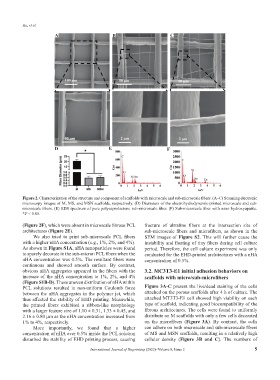
Figure 2. Characterization of the structure and component of scaffolds with microscale and sub-microscale fibers. (A–C) Scanning electronic
microscopy images of M, MS, and MSN scaffolds, respectively. (D) Diameters of the electrohydrodynamic-printed microscale and sub-
microscale fibers. (E) EDS spectrum of pure polycaprolactone sub-microscale fiber. (F) Sub-microscale fiber with nano hydroxyapatite.
*P < 0.05.
(Figure 2F), which were absent in microscale fibrous PCL fracture of ultrafine fibers at the intersection site of
architectures (Figure 2E). sub-microscale fibers and microfibers, as shown in the
We also tried to print sub-microscale PCL fibers SEM images of Figure S2. This will further cause the
with a higher nHA concentration (e.g., 1%, 2%, and 4%). instability and floating of tiny fibers during cell culture
As shown in Figure S1A, nHA nanoparticles were found period. Therefore, the cell culture experiment was only
to sparely decorate in the sub-micron PCL fibers when the conducted for the EHD-printed architectures with a nHA
nHA concentration was 0.5%. The resultant fibers were concentration of 0.5%.
continuous and showed smooth surface. By contrast,
obvious nHA aggregates appeared in the fibers with the 3.2. MC3T3-E1 initial adhesion behaviors on
increase of the nHA concentration to 1%, 2%, and 4% scaffolds with micro/sub-microfibers
(Figure S1B-D). These uneven distribution of nHA within
PCL solutions resulted in non-uniform Coulomb force Figure 3A-C present the live/dead staining of the cells
between the nHA aggregates in the polymer jet, which attached on the porous scaffolds after 4 h of culture. The
thus affected the stability of EHD printing. Meanwhile, attached MT3T3-E1 cell showed high viability on each
the printed fibers exhibited a ribbon-like morphology type of scaffold, indicating good biocompatibility of the
with a larger feature size of 1.10 ± 0.31, 1.33 ± 0.45, and fibrous architectures. The cells were found to uniformly
2.16 ± 0.80 μm as the nHA concentration increased from distribute on M scaffolds with only a few cells decorated
1% to 4%, respectively. on the microfibers (Figure 3A). By contrast, the cells
More importantly, we found that a higher can adhere on both microscale and sub-microscale fibers
concentration of nHA over 0.5% inside the PCL solution of MS and MSN scaffolds, resulting in a relatively high
disturbed the stability of EHD printing process, causing cellular density (Figure 3B and C). The numbers of
International Journal of Bioprinting (2022)–Volume 8, Issue 2 5