
Page 156 - IJB-8-2
P. 156
Application of Bioprinting in Ophthalmology
In this article, we review the main 3D bioprinting in a slower printing speed than the top-down method
techniques, summarize the bioinks adopted in 3D with continuous light scanning. To attain a successful
bioprinting, and discuss the applications of bioprinting printing, it is critical for both methods to have a good
in ophthalmology. We also present the advantages bonding between the printed structure and build-platform.
and limitations of bioprinting in the ocular tissue Immediately after printing, the support structures need to
engineering as well as the future directions that will be removed manually. The advantages of SLA techniques
translate the technologies to personalized therapeutic and include the high lateral and vertical printable resolution
pharmaceutical tools in ophthalmology. (about 20–50 μm and 25–100 μm, respectively) , a
[12]
At present, vat polymerization (VP)-based, material wide range of printable viscosities (up to 5 Pa.s) , high
[13]
extrusion-based, and material jetting-based printing printable cell density (could be 10 cellsper ml), and great
8
strategies are the three principal printing techniques potential to formulate highly complex structures with the
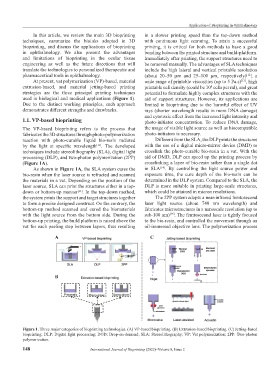
used in biological and medical applications (Figure 1). aid of support structures. However, its applications are
Due to the distinct working principles, each approach limited in bioprinting due to the harmful effect of UV
demonstrates different strengths and drawbacks. rays (shorter wavelength results in more DNA damage)
and cytotoxic effect from the increased light intensity and
1.1. VP-based bioprinting photo-initiator concentration. To reduce DNA damage,
The VP-based bioprinting refers to the process that the usage of visible light source as well as biocompatible
fabricates the 3D structures through photo-polymerization photo-initiators is necessary.
reaction with photo-curable liquid bio-resin radiated Different from the SLA, the DLP prints the structures
by the light at specific wavelength . The developed with the use of a digital micro-mirror device (DMD) to
[11]
techniques include stereolithography (SLA), digital light crosslink the photo-curable bio-resin in a vat. With the
processing (DLP), and two-photon polymerization (2PP) aid of DMD, DLP can speed up the printing process by
(Figure 1A). crosslinking a layer of bio-resin rather than a single dot
As shown in Figure 1A, the SLA system cures the in SLA . By controlling the light source power and
[14]
bio-resin when the laser source is refracted and scanned exposure time, the cure depth of the bio-resin can be
the materials in a vat. Depending on the position of the determined in the DLP system. Compared to the SLA, the
laser source, SLA can print the structures either in a top- DLP is more suitable in printing large-scale structures,
down or bottom-up manner . In the top-down method, which could be attained in micron resolutions.
[11]
the system prints the support and target structures together The 2PP system adopts a near-infrared femtosecond
to form a precise designed construct. On the contrary, the laser light source (about 740 nm wavelength) and
bottom-up method scanned and cured the biomaterials fabricates microstructures in a nanoscale resolution (up to
with the light source from the bottom side. During the sub-100 nm) . The femtosecond laser is tightly focused
[15]
bottom-up printing, the build platform is raised above the to the bio-resin, and controlled the movement through an
vat for each peeling step between layers, thus resulting oil-immersed objective lens. The polymerization process
A C
B
Figure 1. Three major categories of bioprinting technologies. (A) VP-based bioprinting. (B) Extrusion-based bioprinting. (C) Jetting-based
bioprinting. DLP: Digital light processing; DOD: Drop-on-demand; SLA: Stereolithography; VP: Vat polymerization; 2PP: Two-photon
polymerization.
148 International Journal of Bioprinting (2022)–Volume 8, Issue 2