
Page 160 - IJB-8-2
P. 160
Application of Bioprinting in Ophthalmology
the semiconductive polymer onto a hemispherical surface banks against the increasing cases of corneal blindness is
using a customized extrusion printer, Park et al. fabricated the main driver for the development of artificial cornea.
the polymer-based photodetectors that can generate Compared with the traditional technologies for artificial
electricity from the light stimuli . The production of cornea production, 3D bioprinting provides a fast method
[61]
electronic devices by 3D printing confirms that the design to reconstruct individual-specific tissues and organs with
of light receptors is a more flexible and efficient method high reproducibility.
than the traditional manufacturing. Indirectly, this might Bioprinting offers the possibility of producing
facilitate the development of wearable and implantable artificial cornea. The human corneal scanning model
material that can restore the vision in future. is used to print artificial cornea with complex structure
through bioprinting, but the tissue function of artificial
(5) Orbital implant cornea still needs to be further validated in clinical
[31]
3D printing is a flexible and low-cost method for trials . The challenge in bioprinting the cornea lies in the
designing customized complex orbital reconstruction transparency, microporosity and specific shape properties
implants [62-65] . Based on the digital images from the of the structure [74,75] .
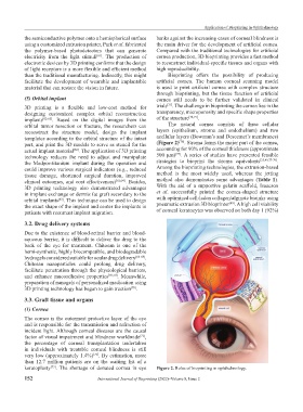
orbital tumor resection or fracture, the researchers can The natural cornea consists of three cellular
reconstruct the structure model, design the implant layers (epithelium, stroma and endothelium) and two
templates according to the orbital structure of the intact acellular layers (Bowman’s and Descemet’s membranes)
[76]
part, and print the 3D models to serve as stencil for the (Figure 2) . Stroma forms the major part of the cornea,
actual implant material . The application of 3D printing accounting for 90% of the corneal thickness (approximate
[62]
[75]
technology reduces the need to adjust and manipulate 500 μm) . A series of studies have presented feasible
the Medpor-titanium implant during the operation and strategies to bioprint the stroma equivalents [35,49,77-79] .
could improve various surgical indicators (e.g., reduced Among the bioprinting technologies, the extrusion-based
tissue damage, shortened surgical duration, improved method is the most widely used, whereas the jetting
clinical outcomes, and cost effectiveness) [62,64] . Besides, method also demonstrates some advantages (Table 1).
3D printing technology also demonstrated advantages With the aid of a supportive gelatin scaffold, Isaacson
in implant exchange or dermis fat graft secondary to the et al. successfully printed the cornea-shaped structure
orbital implants . This technique can be used to design with optimized cell-laden collagen/alginate bioinks using
[63]
[49]
the exact shape of the implant and center the implants in pneumatic extrusion 3D bioprinter . A high cell viability
patients with recurrent implant migration. of corneal keratocytes was observed on both day 1 (92%)
3.2. Drug delivery systems
Due to the existence of blood-retinal barrier and blood-
aqueous barrier, it is difficult to deliver the drug to the
back of the eye for treatment. Chitosan is one of the
hemi-synthetic, highly biocompatible, and biodegradable
hydrogels considered suitable for ocular drug delivery [66-68] .
Chitosan nanoparticles could prolong drug delivery,
facilitate penetration through the physiological barriers,
and enhance mucoadhesive properties [69,70] . Meanwhile,
preparation of nanogels of personalized medication using
3D printing technology has begun to gain traction .
[70]
3.3. Graft tissue and organs
(1) Cornea
The cornea is the outermost protective layer of the eye
and is responsible for the transmission and refraction of
incident light. Although corneal diseases are the causal
factor of visual impairment and blindness worldwide ,
[71]
the percentage of corneal transplantation undertaken
in individuals with treatable corneal blindness is still
very low (approximately 1.4%) . By estimation, more
[72]
than 12.7 million patients are on the waiting list of a
keratoplasty . The shortage of donated cornea in eye Figure 2. Roles of bioprinting in ophthalmology.
[73]
152 International Journal of Bioprinting (2022)–Volume 8, Issue 2