
Page 161 - IJB-8-2
P. 161
Wang, et al.
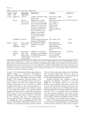
Table 1. Application of 3D bioprinting in ophthalmology
Ocular Tissue Bioprinting Biomaterials Cell types References
tissues layer approaches
Cornea Epithelium Extrusion GelMa bioink/GelMA dome- CEpCs/LECs, Human [50,86]
shaped mold CEpCs line
Stroma Extrusion, Matrigel-COL I bioink/ CSKs/LSSCs, human [28,31,35,49,79,80,86-88]
Inkjet, Laser- LN-COL IV support sheet, LECs+ADSCs,
assisted, SLA ALG-COL I bioink/FRESH human CSKs, rat
support, COL I-AG bioink/ LSSCs, human
no support, GelMa bioink/ TDMSCs with
reinforced with PEG-PCL keratocyte induction,
fibers, GelMA bioink/ no HCKs
support, cornea-derived
dECM bioink/no spport,
ALG/GEL bioink/resin
support
Endothelium Extrusion Gelatin-RGD bioink/amniotic CECs, human CECs [51,86]
membrane dECM support
Retina Retinal Laser-assisted, HA-GM and PEG-RGDs, RPEs, human fetal [44,89]
pigment Microvalve DMEM:F12/ALG and retinal progenitor cells,
epithelium jetting Pluronic ARPE-19, human
retinoblastoma cell line
(Y79)
Retinal Piezoelectric DMEM, ITO-coated glass, Retinal granlion cell [54,83,90]
ganglion inkjet, 2PP, alginate and culture Medium/ neurons, retinal glial
cells thermal inkjet PLA/HEIP and matrigel for cells, human iPSC,
combined with electrospinning retinal ganglion cells
electrospinning
ADSCs: Adipose-derived stem cells; AG: Agarose; ALG: Alginate; CECs: Corneal endothelial cells; ARPE-19: Adult retinal pigmented epithelial cell line-19;
CEpCs: Corneal epithelial cells; COL: Collagen; CSK: Corneal stromal keratocytes; dECM: Decellularized extracellular matrix; DMEM: Dubelcco’s
Modified Eagle’s medium; FRESH: Freeform reversible embedding of suspended hydrogels; GelMA: Gelatin methacrylate; HA-GM: Hyaluronic acid
with methacrylation by glycidyl-hydroxyl reaction; HCKs: Human corneal keratocytes; HEIP: Hexafluoroisopropanol; iPSC: Induced pluripotent stem cell;
ITO: Indium tin oxide; LECs: Limbal epithelial cells; LN: Laminin; LSSCs: Limbal stromal stem cells; PEG-PCL: Polyethylene glycol-polycaprolactone;
PEG-RGDs: Arg-Gly-Asp-Ser peptide; PLA: Polylactic acid; RPE: Retinal pigment epithelial cells; SLA: Stereolithography; TDMSCs: Turbinate-derived
mesenchymal stem cells; 2PP: Two-photon polymerization
and day 7 (83%) post-printing. Without using supportive for the survival and phenotype maintenance of keratocyte
scaffold, Campos et al. adopted an electromagnetic cells. In another study, shear stress was induced by
micro-valve, rather than micro-extrusion, to directly print printing nozzles of different sizes in order to organize the
the corneal structure with collagen hydrogel in a DOD printed collagen fibrils in the lattice pattern .
[79]
manner . The transparency and optical density of the Besides, researchers also attempted to print the
[77]
printed structure was comparable to those of the native endothelial and epithelial components of the cornea
cornea, and the keratocyte cells assumed the typical (Table 1) . For instance, Sorkio et al. printed the
[45]
dendritic morphology on day 7 post-printing. GelMA stratified corneal epithelium and stroma construct with
is another good option for stroma printing; however, in the stem cell-laden laminin/collagen bioink using laser-
GelMA-only printed structure, the keratocytes stayed assisted technology . Jin et al. printed the cells of corneal
[28]
in round shapes after days . The specially designed epithelium using DLP technology coupled with extrusion
[35]
alignment of collagen fibrils is critical to the transparency printing . Kim et al. printed the corneal endothelium
[50]
of the cornea. To mimic the sophisticated topological cells using extrusion printing . The results of these
[51]
alignment of the stroma structure, Kong et al. fabricated studies strengthened the possibility that corneal substitute
the 3D construct with poly (ε-caprolactone)-poly can be rapidly generated. Nevertheless, further in vitro
(ethylene glycol) (PECL) microfibrous scaffold, which and vivo studies are still needed for clinical validation.
is a modified type of poly (ε-caprolactone) (PCL) with Hence, 3D bioprinting technologies embody a great
improved hydrophilicity, and GelMA hydrogel by direct potential and show promising prospects in the fabrication
writing . The construction provided a good environment of artificial cornea. Efforts have been made to improve
[78]
International Journal of Bioprinting (2022)–Volume 8, Issue 2 153