
Page 272 - IJB-8-3
P. 272
Bone Sialoprotein enhances Bone Regeneration
A growing along and inside the PLA cylinder increase in the
groups with both growth factors. In the group with BSP,
most bone areas are detected around the cylinder and only
marginally growing into the cylinder, in the group with
BMP-7 growth inside the cylinder was detected and only
marginal growth outside the cylinder. These observations
are confirmed by quantitative analyses where significant
differences are determined regarding area of new formed
bone between the control group with collagen alone
and the groups with high BSP and BMP7. Interestingly,
no significant differences are observed between the
groups BSP and BMP-7.
BMP-7 has been used as control as its effects
on bone formation have been described in detail .
[50]
However, administration of BMPs (either BMP-2 or
BMP-7) can result in many side effects such as excessive
bone formation , heterotopic ossification , bone
[51]
[52]
with atypical structure or low mechanical stability ,
[53]
inflammatory complications or massive soft-tissue
swelling, or tumor formation .
[54]
By reflecting former results regarding BSP
immobilized on titanium or CPC [11,12,14,15] and comparing
them with the results of the presented study, we confirmed
our hypothesis that the best effects of BSP are achieved
when BSP is coupled to collagen type I. It is known that
BSP contains a collagen-binding site and several effects
[19]
regarding the interaction between collagen and BSP have
B been described. Choi et al. showed that the collagen
interaction promoted hydroxyapatite nucleation and that
a collagen-binding peptide derived from BSP increased
osteogenic differentiation in muscle-derived stem cells
and induced expression of osteoblastic marker genes
and proteins without affecting proliferation . Last but
[55]
not least, Choi et al. showed coating of hydroxyapatite
scaffolds with the collagen-binding peptide-induced bone
formation in a craniotomy defect in rabbits . Similar
[56]
effects were observed in a rat calvarial model [20,21] , where
they found induced calcification. Comparing their studies
with ours, they used higher concentrations than we did
(20 μg/implant). Moreover, they did not show any data
on bone regeneration by X-rays or μCt analyses, but
only histological pictures after 30 days, which makes an
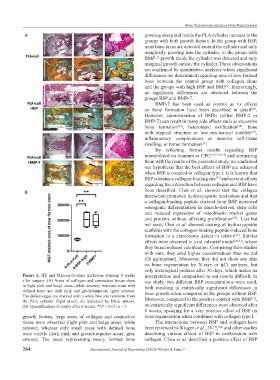
Figure 6. HE and Masson-Goldner trichrome staining 8 weeks interpretation and comparison to our results difficult. In
after surgery. (A) Areas of collagen and connective tissue show our study, two different BSP concentrations were used,
in light pink and beige areas (white arrows), whereas areas with both resulting in statistically significant differences in
defined bone are dark pink and green/turquoise (gray arrows).
The defect edges are marked with a white line and remnants from bone growth when compared to the groups without BSP.
the PLA cylinder (light areas) are indicated by black arrows. Moreover, compared to the positive control with BMP-7,
(B) Quantification of results after 8 weeks. **P < 0.01; n = 5. no statistically significant differences were observed after
8 weeks, speaking for a very positive effect of BSP on
growth factors, large areas of collagen and connective bone regeneration when combined with collagen type I.
tissue were observed (light pink and beige areas; white The interactions between BSP and collagen have
arrows), whereas only small areas with defined bone been reviewed by Kruger et al., 2013, and other studies
[22]
were visible (dark pink and green/turquoise areas; gray describing various effects of BSP in combination with
arrows). The areas representing newly formed bone collagen. Chou et al. described a positive effect of BSP
264 International Journal of Bioprinting (2022)–Volume 8, Issue 3