
Page 269 - IJB-8-3
P. 269
Kriegel, et al.
A
B
C D
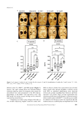
Figure 3. µCt images 3 weeks (A) and 8 weeks (B) after surgery. (C and D) Quantification of results after 3 and 8 weeks. *P < 0.05,
**P < 0.01, ***P < 0.005; ****P < 0.001 (n = 5).
defects in the CG, BMP-7, and BSP groups (Figure 3, BSP, we observe a better bone regeneration. In a calvarial
arrows). The right column shows the Masson-Goldner defect model with calcium phosphate scaffolds coated
trichrome staining, where the turquoise stain stands for with BSP, no statistical significances were observed
mineralized bone and corresponds to the HE staining. between the groups regarding bone thickness and bone
Interestingly, in the BMP-7 and especially in the 5 μg fraction . No positive effect on bone regeneration was
[14]
BSP group, an intense orange staining can be observed observed with the same scaffolds used in a femoral condyle
representing osteoid tissue. defect model coated with BSP . Considering our results
[15]
Comparing our results using collagen with other in in relation to the literature lead to the hypothesis that the
vivo models employing implant materials coated with material used as a scaffold plays an important role for the
International Journal of Bioprinting (2022)–Volume 8, Issue 3 261