
Page 270 - IJB-8-3
P. 270
Bone Sialoprotein enhances Bone Regeneration
and collagen type I as biodegradable polymer material,
which can easily be modified [26,27] . In vitro analyses of
this material were performed and demonstrated a good
biocompatibility . As described in the method section,
[27]
we printed a 3D cylinder of polylactide which was
filled manually with collagen type I and two different
concentrations of BSP. Binding between BSP and collagen
type I is provided by the collagen type I binding site of
BSP, which has been described in several studies [18,19,22] .
BSP release was measured as described in materials
and methods and is shown in Figure S3. We observed a
steady release of BSP over a time period of 72 h. After
this time, approximately 60% of the immobilized protein
was released, which complies with former studies [26,27] .
The dose of 100 ng/ml has been used in various studies.
Furthermore, the slow release has been described and
is likely to be beneficial for osteogenic regeneration .
[48]
Our hypothesis is that the missing 40% still remain in the
gel and are released in a slow manner, however, further
studies with longer time points and determination of
the residue dosage have to be performed to confirm this
theory.
PLA cylinders were modified with collagen, BSP,
or BMP-7 and implanted into a femoral defect of rats.
X-rays performed every 2 week demonstrated the
nd
course of bone healing. Figure 5A (exemplary images)
shows that in the groups with growth factors, either BSP
in two concentrations or with BMP-7, the bone defect
is almost closed 8 weeks after surgery. Concerning the
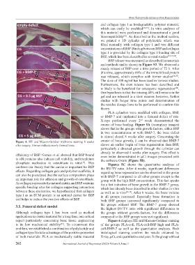
Figure 4. HE and Masson-Goldner trichrome staining 8 weeks course of bone growth, it can be observed that BMP-7
after surgery. Arrows indicate newly formed bone. shows an earlier begin of bone regeneration than BSP,
particularly a directed growth through the cylinder can
already be observed 4 weeks after surgery. This effect is
efficiency of BSP. Gomes et al. showed that BSP bound even better demonstrated in μCt images processed with
to silk proteins also induces cell viability, and tricalcium the software Osirix (Figure 5B).
phosphate nucleation in osteoblasts in vitro . This Figures 5C shows the quantitative analyses of
[13]
confirms our theory that the carrier is important for BSP the BV/TV ratio. After 4 weeks, significant differences
effects. Regarding collagen gels and polymer scaffolds, it regarding bone regeneration can be observed in the group
can also be postulated that the surface composition plays with BMP-7 compared to all other groups except to the
an important role for adhesion and growth of osteoblasts. group with the high BSP concentration. This fact speaks
As collagen represents the natural matrix and BSP contains for a fast induction of bone growth in the BMP-7 group,
specific binding sites for collagen supporting interaction which has already been described in other studies in vitro
between these molecules, we hypothesized that collagen as well as in vivo [26,49] . After 8 weeks, the bone volume
type I as an ECM protein is an optimal carrier for BSP in all groups increased. Especially bone formation in
and helps to induce the positive effects of BSP. both BSP groups increased significantly compared to
3.5. Femoral defect model the groups without BSP. The BMP-7 group showed
the highest BV/TV ratio with significant differences to
Although collagen type I has been used in medical the groups without growth factors, but the differences
applications as carrier material for a long time, one critical compared to the BSP groups were not significant.
aspect particularly concerning bone tissue engineering Figure 6 displays HE and Masson-Goldner staining
is its low mechanical stability . To circumvent this of the groups PLA-coll, PLA-coll-BSP high and PLA-
[47]
problem, we established a combination of polylactide and coll-BMP-7 as well as the quantitative analyses. Both
collagen type I to take advantage of the positive properties histological staining confirm the results obtained by
of both materials: PLA as mechanically stable material X-ray, μCt, and quantitative analyses. In the group without
262 International Journal of Bioprinting (2022)–Volume 8, Issue 3