
Page 15 - IJB-8-4
P. 15
Shi, et al.
A
B
C
D
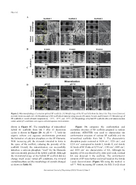
Figure 2. Micromorphology of extrusion-printed SF scaffolds. (A) Morphology of the SF scaffolds from the three inks: Top views (first row)
and side views (second row). (B) Morphology of SF scaffolds at varied printing speeds of 6 mm/s, 8 mm/s, and 10 mm/s. (C) Morphology of
SF scaffolds at varied substrate temperatures, −10°C, −13°C, and −15°C. (D) Morphology of hybrid SF scaffolds after post-mineralization
under varied pH conditions and organic solvent/aqueous environments.
shown in Figure 1C. The morphology of mineralized Figure 3A compares the conformation and
hybrid SF scaffolds from Ink 3 after 15 deposition crystalline structure of SF scaffolds prepared in various
cycles is shown in Figure 2D. At pH 4 – 5, both the conditions. ATR-FTIR was used to characterize the
organic solvent and aqueous environments promoted conformation structure of various SF scaffolds and the
the formation of calcium phosphate on the SF filaments. mineralized scaffolds from Ink 3. The characteristic
With increasing pH, the minerals intended to form in absorption peaks centered at 1624 cm , 1526 cm , and
-1
-1
the space of the scaffold, reducing the porosity of the 1233 cm correspond to Amide I, Amide II, and Amide
-1
scaffold. Overall, the mineralization can successfully III bands of SF. Peaks at 3279 cm , 1590 cm , 1409 cm ,
-1
-1
-1
introduce a calcium phosphate “shell” for the filaments and 1030 cm are characteristic of SA. Although the
-1
and conveniently produced the hybrid scaffold. Although majority of SA was dissolved by citric acid wash, residual
the micromorphology of the hybrid SF scaffold did not SA was detected in the scaffolds. The conformation
change much under varied pH conditions, the mineral contents of SF were further analyzed based on the Amide
crystalline phase and the morphology of crystals changed I peak deconvolution (Figure S3) using the method in
as shown in Table S1. ref. . With increasing SF content, the Silk II or β–sheet
[41]
International Journal of Bioprinting (2022)–Volume 8, Issue 4 7