
Page 19 - IJB-8-4
P. 19
Shi, et al.
A B C
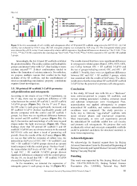
Figure 5. In vitro assessments of cell viability and osteogenesis effect of 3D printed SF scaffolds using osteocytes MC3T3-E1. (A) Cell
viability was evaluated by CCK-8 assay. (B) Cell osteogenic property was evaluated by ALP assay. (C) The osteogenesis-related genes
were detected by q-PCR on day 10 and compared by relative mRNA expression. Significant differences are denoted as: * for P<0.05, ** for
P<0.01, *** for P<0.001 compared to the control group; for P<0.05, for P<0.01, ### for P<0.001 compared to OIC and OIC+SF-scaffold
#
##
3 groups.
Interestingly, the Ink 2-based SF scaffolds exhibited The results showed that there were significant differences
the greatest modulus. The acidic residues and hydrophilic in osteogenesis-related genes (Runx2, OPN, OCN, OSX,
groups can interact better with Ca , thus leading to more and Col1a) between OIC + SF scaffold 3-CaP10 and
2+
calcium mineral [53,54] . β-sheet conformation tended to the rest of groups including control, OIC, and OIC + SF
induce the formation of calcium minerals. Nevertheless, scaffold 3. Notably, there were no significant differences
we propose multiple reasons that resulted in the high between OIC and OIC + SF scaffold 3 groups, which
modulus of the SF scaffolds, and the establishment of was consistent with the results of ALP assay. The above
structure-morphology-mechanical property correlations results prove that the mineralized SF scaffold (SF scaffold
requires future investigations. 3-CaP10) has the potential to promote cell osteogenesis.
3.5. 3D printed SF scaffold 3-CaP10 promotes Conclusion
cell proliferation and osteogenesis
In this study, SF-based inks with SA as a “thickener”
According to the results of our CCK-8 experiment, on were extrusion-printed to prepare 3D scaffolds, and
the 1 day, there was no significant difference of OD various printing parameters including extrusion speed
st
value between the control, SF scaffold 3, and SF scaffold and substrate temperature were investigated. Post-
3-CaP10 groups (Figure 5A). On the 3 and 5 days, mineralization was applied subsequently to prepare
rd
th
the OD value in each group significantly increased, and mineralized SF scaffolds, and various mineralization
SF scaffold 3-CaP10 showed significantly greater cell conditions were compared. The study provides a
density compared with the control and SF scaffold 3 facile way to fabricate “egg-shelled” scaffolds with
groups, but there was no significant difference between tuned mineral phases and mechanical properties.
the control and SF scaffold 3 groups (Figure 5A). On Most importantly, in vitro cell experiments proved
day 5, the number and morphology of cells were in good the mineralized SF scaffolds exhibit low cell toxicity
shapes under the light microscope (Figure S5). These and promote cell osteogenesis. We propose that such
findings show that 3D printed SF scaffold 3 and SF mechanically robust and osteocyte-compatible scaffolds
scaffold 3-CaP10 have no obvious toxicity to the normal could be potential candidates for structural materials in
MC3T3-E1 cells and show a trend of promoting cell bone tissue engineering.
growth. From the results of the ALP assay (Figure 5B),
we can see that with the extension of culture time, the Acknowledgments
intracellular ALP level of OIC, OIC + SF scaffold 3, and
OIC + SF scaffold 3-CaP10 groups showed an upward We acknowledge the financial support from the Beijing
trend compared with the control group. On days 4 and 7, Advanced Innovation Center for Biomedical Engineering of
the intracellular ALP levels of OIC, OIC + SF scaffold Beihang University and Capital Medical University Affiliated
3, and OIC + SF scaffold 3-CaP10 groups showed Beijing Chaoyang Hospital.
significant differences compared with the control group. Funding
On day 10, we found that OIC + SF scaffold 3-CaP10
showed the highest ALP level. This work was supported by Wu Jieping Medical Fund
In the meantime, we detected the expression of (No. 320.6750.2020-06-12), Beijing Natural Science
osteogenic genes in each group on day 10 (Figure 5C). Foundation (No. L202006), National Key R&D
International Journal of Bioprinting (2022)–Volume 8, Issue 4 11