
Page 200 - IJB-8-4
P. 200
A Review on Bioinks and their Application in Plant Bioprinting
gels (DM 50%) are produced by the production of extensively utilized in food-based formulations, some
calcium crosslinks between free carboxyl groups [144] . chemicals used in this composition were not consumable.
The gelation mechanism of LM pectin is comparable to As a result, “bioink” is a more suitable name than “food-
that of alginate with calcium, known as the “egg-box” ink” [141] .The color of the printed cubes changed depending
model of gelation [145] . Lamb’s lettuce cells (Valerianella on the bioink used; if the ink contains only pectin gel and
locusta) were selected as the plant cells for encapsulation no cells, the printed cubes will have a yellowish hue, while
into pectin-based bioinks. Before isolating cells, a bioinks with lettuce cells usually produce a green hue,
precise technique was followed, that is, Lamb’s lettuce due to the presence of chloroplasts in the plant cells [141] .
(V. locusta, (L) Laterr., var. “Gala”) was collected early Lightly-colored printed cubes usually were a product of
in the morning from a commercial greenhouse under a the presence of BSA, which regulates the existence of air
12 h light/dark regime at 150 μm Em s . To remove bubbles that induced transmission of light [147] .
−1
−2
dirt debris, the Lamb’s lettuce was cleaned with chlorine This method helps encapsulate land plant cells
distilled water prior to shipment [146] . The purchased in high-density pectin gels with greater efficacy and
Lamb’s lettuce was washed with 0.0005% NaOCl, rinsed consistency than previous methods [141] . To date, 8 pectin-
5 times with deionized water, and then dried before use. based bioinks, with and without cells, have been produced
The pectin solution was made using methoxylated using this method. The framework introduced in this
pectin extracted from the peel of citrus and calcium research can be viewed as the first step toward producing
chloride dihydrate [141] . 3D-printed particulate or cellular foods.
D-glucose anhydrous, 4-morpholine ethane sulfonic
acid hydrate, magnesium sulfate heptahydrate, bovine 6.2. Synthetic bioink
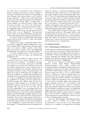
serum albumin (BSA), and pectinase from Aspergillus 6.2.1. 3D bioprinting of artificial leaves
niger were used as buffer solutions. Evans blue,
fluorescein diacetate, Hoechst 33258, and pentahydrate For the reproduction of the primary structural features of a
(bisbenzimide) were employed to determine viability [141] . leaf for quick mass transmission, 3D hierarchical macro/
All pectin solutions used in this process were mesoporous structures with high surface areas must be
prepared the day before the lettuce cell extraction and synthesized [148] . Titanium dioxide-based inks are ideal for
maintained at 4°C. creating 3D-printed leaf structures with high structural
To prepare the bioink, the pectin solution with integrity and surface area as well as porosity-tunable 3D
encapsulated cells and a CaCl suspension (in a 1:1 hierarchical constructions [149] (Figure 16).
2
ratio) were mixed together [141] . To modify the porosity The ink was prepared by producing a sol-gel
and viscosity of the bioink, varied pectin and BSA precursor solution called titanium diisopropoxide
concentrations were used to make 8 distinct bioinks with bis(acetylacetonate) (TIA), which contains two
and without embedded lettuce cells. In total, 20 mL of isopropoxide radicals surrounding one titanium atom, two
pectin was prepared for this experiment. Before the final acetylacetone groups, as well as dodecyl benzenesulfonic
preparation of solution, 10 mL of the pectin solution was acid (DBSA), which helps to improve the surface area.
−1
3
stirred for 10 min at 10,000 rpm, and air bubbles were The pore volume was set to 0.1 cm g , and the DBSA
stabilized using BSA. The solution was then agitated with content is determined to obtain an optimal surface area
a magnetic stirrer set at high speed. Lettuce cell suspension consistent with that of typical mesoporous structures.
(2 × 10 mL) was obtained by decanting 2 falcon tubes of However, producing TIA-based bioinks is often difficult
raw suspension and washing with CaCl solution (26 or because of diluted TIA suspensions, which are classified
2
40 mM) overnight. The viability of the cell and CaCl as low-viscosity Newtonian fluids, thus lacking the
2
suspensions, which contained 13 or 20 mM CaCl , was requisite rheological behavior for 3D printing [150] . The
2
determined using the Evans Blue Exclusion staining DBSA-TIA ink should always sustain its cylindrical
procedure after decanting for 20 min. Then, the cell shape after ejection through a micronozzle, and it should
and CaCl suspensions (5 mL of each falcon tube) were have the capacity to span across spaces over underlying
2
added dropwise to the pectin solution. When the solution surfaces for large-scale fabrication of 3D structures [151-153] .
became homogeneous, a gel formed almost immediately. DBSA-TIA-based designs contain numerous woodpile
Finally, the prepared bioink was lightly stirred for 5 min layers, each of which is orthogonal to the one before
with a spatula, after which the bioink could be used for it and connected by printed filaments spanning spaces
bioprinting. The viscosity and porousness of the ink can between underlying surface structures.
be adjusted by altering the pectin and BSA concentrations There were other methods of bioprinting structures
of the ink precursor, and the cell survival was unaffected of micro leaves [150] . Notably, the artificial micro
by the formulation of pectin-based bioink processing leaf structure is similar to that of the macroporous
and printing [141] . Although pectin-based biomaterials are architecture of a natural leaf, implying the possibility
192 International Journal of Bioprinting (2022)–Volume 8, Issue 4