
Page 203 - IJB-8-4
P. 203
Ghosh and Yi
augmentation in algal biotechnology [159,160] . Furthermore, Membrane fabrication was the following phase,
this type of setup can aid the investigation of natural coral wherein silicone elastomer polydimethylsiloxane
morphology and incite translation to other contexts. (PDMS) was combined with the curing agent at a 10:1
ratio and poured on top of the PMMA mold. The PDMS
6.3.2. 3D printing of leaf-like structures for CO2 duplicate was peeled off from the PMMA mold for the
reduction suggested artificial leaf’s bottom reservoir after being
Liu et al. were the first use cyanobacterial metabolism hardened in an oven. The spin speed was carefully
to recover CO in a hierarchically porous and transparent managed to establish a 40 μm gas-permeable layer on
2
microsystem [165] . A 3D architecture of a natural leaf top of the PDMS membrane [166] . Using a laser cutting
with multi-scaled levels was imitated so that (i) water/ tool, the botanic fiber layer was meticulously designed.
nutrients could be transmitted to the bacterial cells Due to evaporation from sunlight, nutrients and water
through the botanic fiber layer (c.f. xylem and phloem), were transmitted from an external media reservoir to
(ii) the cyanobacterial layer in the system could perform photosynthetic bacterial cells, while capillary force
photosynthesis and respiration to decrease CO levels (c.f. pushed the botanic fiber tube along, imitating the xylem
2
mesophyll), and (iii) solar evaporation could help build and phloem of a natural leaf [165].
up capillary force through the translucent and porous The sol-gel transition of the bioink (i.e., hydrogel-
membrane layer (c.f. epidermis), potentially allowing encapsulated cyanobacteria) must be carefully controlled
self-sustaining capabilities. The authors developed a for materials to retain their shape when patterned on
self-sustaining, biological artificial leaf that significantly the botanic fiber layer in this bioprinting method. In
lowered CO levels in the atmosphere and exchanged it deionized water with 0.5 M CaCl , cyanobacterial cells
2
with O 2 [165] . Moreover, this artificial leaf structure can aid were encased in 6% (w/v) alginate. 2
in understanding the natural leaf structure, water, and gas The diffusion time required for the sol-gel transition
transport processes within natural leaves. increased as the calcium concentration increased, resulting
The preparation started with the culture of bacterial in greater shear stress on the cells. As the rate of the
inoculum. The cyanobacterial stain Synechocystis sp. PCC reaction was slow, a partial uncross linked alginate layer
6803 was utilized for this procedure. Synechocystis spp. could be deposited on the crosslinked layer; this bioink
PCC 6803 was grown from glycerol stock cultures at could be used to print several multilayered patterns. The
80°C by inoculating 15 mL of BG-11 medium under printing was conducted using a 3D Vitarix™ bioprinter
5
gentle shaking with 12 h light and dark intervals. The BG- with the following settings: pressure: ~20 Mkpa; print
11 media consisted of 40 mg of K HPO , 1.5 g of NaNO , speed: 25 mm s , infill density: 30%; and nozzle size:
−1
4
2
3
36 mg of CaCl , 75 mg of MgSO , 6 mg of citric acid, 23 Gauge.
2
4
1 mg of EDTA, and 6 mg of ferric ammonium citrate per The final stage involved the bioprinting of the
1 L of distilled water. For 2 weeks, a fluorescent lamp- cyanobacterial cell-laden hydrogel on top of the botanic
controlled chamber offered continuous aeration and fiber layer and covering it with a gas-permeable PDMS
illumination at a temperature of 30 ± 2°C. The optical membrane. The system generated a hybrid hierarchical
density at 600 nm (OD ) was used to track growth. cell/alginate architecture, with the gas permeable PDMS
600
5 BG11 is a universal medium for the cultivation and maintenance of blue membrane encouraging gas exchange to the bacteria and
green algae (cyanobacteria). botanic fiber layer supplying nutrients and water.
A D E
B C F G
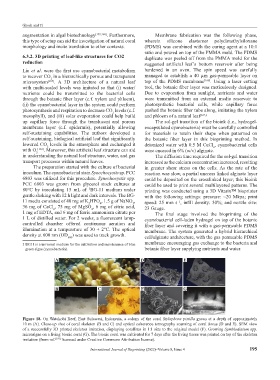
Figure 18. On Watakobi Reef, East Sulawesi, Indonesia, a colony of the coral Stylophora pistilla grows at a depth of approximately
10 m (A). Close-up shot of coral skeleton (B and C) and optical coherence tomography scanning of coral tissue (D and E). SEM view
of a successfully 3D printed skeleton imitation, displaying corallites in 1:1 size to the original model (F). Growing Symbiodinium spp.
microalgae on a living bionic coral (G). The bionic coral was cultivated for 7 days after the living tissue was printed on top of the skeleton
imitation (from ref. [159] licensed under Creative Commons Attribution license).
International Journal of Bioprinting (2022)–Volume 8, Issue 4 195