
Page 287 - IJB-8-4
P. 287
Fayyazbakhsh, et al.
2.8.2. Wound closure with an Olympus DP70 digital camera) using the ×4
Dermal wounds were photographed every week after and ×10 objective lenses. The entire tissue section was
removing the old dressing and before rebandaging, to scanned, digitally photographed, and “photomerged” to
track the wound size, color, necrotic tissue formation, form a single composite image using Adobe Photoshop
and any trauma caused by dressing removal. A sterile (Adobe, CA, USA). Quantitative histomorphometry was
disposable ruler was inserted close to the wound as the performed by measuring the epidermal layer, dermal
scale. The wound size was quantified by tracing the layer, and granulation tissue (GT) thickness. H&E-stained
wound border in each photograph using ImageJ software. sections were blindly graded by two trained graders with
The wound closure was calculated as follows: sections scored on a scale of 0 – 4 in terms of ER, dermal
regeneration (DR), and GT thickness , as depicted in
[33]
A − A
Wound closure()% = 0 t ×100 (5) Table 2. ImageJ and Adobe Photoshop software were
A used for histomorphometric evaluations and tissue slide
0
photo merging.
where A is the wound area immediately after wound
0
implementation, and A is the wound area at time t (i.e., 1, 2.9. Statistical analysis
t
2, 3, and 4 weeks). Traumatic removal was evaluated by
assessing the presence of traumatic laceration, bleeding, All experimental results were reported as the means ±
and redness in wound margins and surrounding tissues SD of at least three replications for each sample per test.
after the dressing removal. Statistical significance was determined using one-way
ANOVA and Student’s t-test with (P < 0.05) as the level
2.8.3. Histology analysis of significance.
Wound tissue explants (25 × 25 mm ) were resected 3. Results and discussion
2
and fixed overnight in 10% neutral buffered formalin,
then cut into 1 mm thickness tissue blocks that include 3.1. Rheological behavior and shear thinning
wound tissues, margins, and surrounding tissues. Tissue behavior
blocks were processed and embedded in paraffin using
a fully automated tissue processor (TissueTek 2000, An important question associated with hydrogel 3D
Sakura Finetek, CA, USA). Tissue blocks were sectioned printing is the effect of rheological properties on
at 5 μm and mounted on positively charged glass slides printability. Printable materials ideally enable the
for staining with hematoxylin and eosin (H&E). The consistent flow and reproducible fabrication. Viscosity
slides were imaged using a transmitted light bright has been widely reported as the determinant factor
field microscope (Olympus BX53 microscope fitted of printability in viscoelastic materials, for example,
A B C
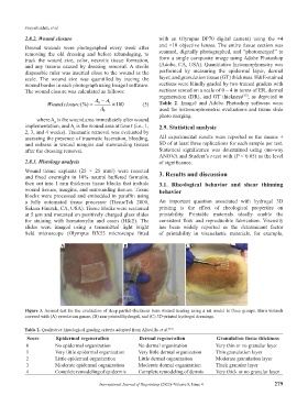
Figure 3. Animal test for the evaluation of deep partial-thickness burn wound healing using a rat model in three groups. Burn wounds
covered with (A) petrolatum gauze, (B) non-printed hydrogel, and (C) 3D-printed hydrogel dressings.
Table 2. Qualitative histological grading criteria adopted from Altavilla et al. [33]
Score Epidermal regeneration Dermal regeneration Granulation tissue thickness
0 No epidermal organization No dermal organization Very thin or no granular layer
1 Very little epidermal organization Very little dermal organization Thin granulation layer
2 Little epidermal organization Little dermal organization Moderate granulation layer
3 Moderate epidermal organization Moderate dermal organization Thick granular layer
4 Complete remodeling of epidermis Complete remodeling of dermis Very thick or no granular layer
International Journal of Bioprinting (2022)–Volume 8, Issue 4 279