Page 292 - IJB-8-4
P. 292
3D Printed Dressings for Burn Wound Treatment
           and G4-A4 dressings. The lower free volume and higher   to the ECM. Gelatin contains RGD peptide sequences,
           entanglement are associated with higher consistency and   which are essential for stable communication between the
           flowability. It also justifies the lower viscosity and better   cells and the surrounding ECM. The presence of amino
           shape  fidelity  outputs  in  G4-A4  and  G6-A2  dressings,   acids and RGDs in the gelatin network provides favorable
           which have the most entanglement between the gelatin   anchors for cell attachment and proliferation to enhance
           amide groups and alginate carboxylate/hydroxyl groups.   cell  adhesion through interactions with integrin  and
           These results collectively  indicate  the promising burn   fibronectin [50,51] . On day 7, 3D-printed G6-A2 dressings
           wound healing capacity of the G6-A2 dressing.       showed higher cell viability than the control group and
                                                               other groups, which confirms the long-term cell viability
           3.5. In vitro biological evaluation                 of the G6-A2 dressings.
           The  in vitro  biocompatibility of 3D-printed gelatin-  The Live/Dead assay results in Figure 11 indicate
           alginate dressings was evaluated by MTT assay and Live/  only a  few dead  cells  in  G6-A2 cell-laden  dressings,
           Dead assay, using HDFs, as depicted in Figures 10 and   associated  with  higher  RGD available  in  this  dressing.
           11. The viability and proliferation of HDF cells after 24 h   However, this sample showed no significant difference in
           of  exposure  to  the  dressings  extracts  were  determined   cell survival compared to the control group (P > 0.05). In
           using an MTT assay after 1, 3, and 7 days to measure OD   contrast, it showed significantly higher cell viability and
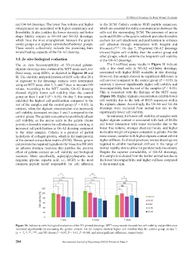
           values. According to the MTT results, G6-A2 dressing   biocompatibility than the rest of the samples (P < 0.05).
           showed slightly lower cell viability  than the control   This  is  consistent  with  the  findings  of  the  MTT  assay
           group on days 1 and 3 (P > 0.05). On day 7, this sample   (Figure 10). Higher alginate concentration exhibits lower
           exhibited the highest cell proliferation compared to the   cell viability due to the lack of RGD sequences within
           rest of the samples and the control group (P < 0.05). In   the alginate chains. Accordingly, the G0-A8 and G2-A6
           contrast, when the alginate concentration was increased,   dressings  were  excluded  from  animal  test  due  to  the
           cell viability decreased on days 1 and 3 compared to the   significantly lower cell viability.
           control group. The gelatin concentration positively affects   In summary, the lower cell viability of samples with
           cell  viability, as the  amino  acids in  the  gelatin  chains   higher alginate content is associated with lack of RGDs
           provide a favorable matrix for cell attachment, resulting in   and lower interaction  with water molecules  due to the
           increased cell proliferation in G6-A2 dressing compared   lower free volume, stronger chemical bonds, and higher
           to the other samples. Gelatin  is a product of partial   molecular weight of alginate compared to gelatin. For the
           hydrolysis of collagen protein, which is the main protein   same reason, samples with higher alginate content exhibit
           of the dermal extracellular matrix (ECM). Hence, gelatin   higher stiffness. In clinical practices, wound dressings are
           can provide the required ingredients for bioactive DR with   required to exhibit mechanical stiffness in the range of
           no  adverse  immune  response  that  justifies  the  positive   normal healthy skin to allow for painless body movement.
           effect of gelatin content on cell viability and biological   Despite  the  superior  extrudability  of  G4-A4  dressings,
           response.  More  specifically,  arginylglycylaspartic  acid   this sample is excluded from the further animal test due to
           (arginine–glycine–aspartic  acid,  i.e.,  RGD) is  the  most   the lower biocompatibility and higher stiffness compared
           common  peptide  motif  responsible for cell  adhesion   to the normal skin.


           Figure 10. Indirect in vitro biological evaluation of the 3D-printed dressings. MTT assay results showed that cell viability and proliferation
           increased significantly by increasing the gelatin content. G6-A2 samples showed higher cell viability than the control group on day 7.
           (n  =  3; *, **, ***, and NS denote P < 0.05, P < 0.01, P <0.001, and non-significant difference, respectively).


           284                         International Journal of Bioprinting (2022)–Volume 8, Issue 4

