
Page 295 - IJB-8-4
P. 295
Fayyazbakhsh, et al.
A
B
C
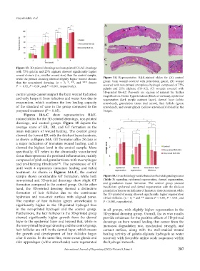
Figure 13. 3D-printed dressings and non-printed G6-A2 dressings
with 75% gelatin and 25% alginate showed significantly higher
wound closure (i.e., smaller wound size) than the control sample,
while the printed dressing showed slightly higher wound closure Figure 14. Representative H&E-stained slides for (A) control
than the non-printed dressing. (n = 3; *, **, and *** denote group: burn wound covered with petrolatum gauze, (B) wound
P < 0.05, P < 0.01, and P < 0.001, respectively). covered with non-printed amorphous hydrogel composed of 75%
gelatin and 25% alginate (G6-A2), (C) wounds covered with
control group cannot support the burn wound hydration 3D-printed G6-A2. Pop-outs are regions of interest for further
magnification. Guide: hyperkeratosis (black arrowhead), epidermal
and only keeps it from infection and water loss due to regeneration (dark purple outmost layer), dermal layer (white
evaporation, which confirms the low healing capacity arrowhead), granulation tissue (red arrow), hair follicle (green
of the standard of care in the group compared to the arrowhead), and sweat glands (yellow arrowhead) labeled in the
proposed treatment (P < 0.05). images.
Figures 14A-C show representative H&E-
stained slides for the 3D-printed dressings, non-printed
dressings, and control groups. Figure 15 depicts the
average score of ER, DR, and GT formation as the
main indicators of wound healing. The control group
showed the lowest ER with the thickest hyperkeratosis,
as shown in Figure 14A. GT formation after 28 days is
a major indication of immature wound healing, and it
showed the highest level in the control sample. More
specifically, GT refers to the chronically vascularized
tissue that represents the persisted inflammation, mainly
composed of pink and granular tissue with macrophages
and proliferating fibroblasts [54] . The persistence of GT
until week 4 represents immature healing and failed
treatment. As shown in Figures 14A-C, the control
sample shows considerable GT formation, while both Figure 15. Gross histology results based on the H&E grading scores
non-printed and 3D-printed dressings show slight GT (Table 3) regarding epidermal regeneration, dermal regeneration,
formation compared to the control group. On the other and granulation tissue formation. The control group showed
hand, the 3D-printed dressing showed a distinctive insufficient epidermal and dermal regeneration with the thickest
granulation layer as an indicator of immature tissue treatment, while
formation of hair follicles due to the continuous the 3D-printed dressing showed significantly higher regeneration
hydration and non-stick surface with aligned pores. of hair follicles. (n = 6; * and ** denote P < 0.05, P < 0.01, and
The number of hair follicles (green arrowheads) is P < 0.001, respectively).
significantly higher in the 3D-printed hydrogel than
in the non-printed hydrogel and the control group. in all groups, with slightly higher regeneration in the
Furthermore, the hair follicles in the 3D-printed group 3D-printed dressing group. Overall, the in vivo results
showed significantly higher growth from the dermal provide evidences for the positive effects of 3D-printed
layer to the epidermal layer and beyond that, while in dressings on burn wound healing that comes from the
the non-printed hydrogel dressing and the control group, increased degradation rate, mechanical strength, and
hair follicles are still in the dermal layer, which means contact surface, along with the well-studied wound
the growth and development of hair follicles began healing activity of gelatin-alginate hydrogels as water
after 4 weeks. In the same line, more sweat glands and reservoir with favorable amino acids sequences within
skin appendages (white arrowheads) were regenerated the hydrogel network.
International Journal of Bioprinting (2022)–Volume 8, Issue 4 287