
Page 284 - IJB-8-4
P. 284
3D Printed Dressings for Burn Wound Treatment
penicillin/streptomycin (pen/strep), MTT, and trypsin/ 2.3. 3D printing
EDTA were purchased from Sigma-Aldrich (St. Louis, We adopted the extrusion-based 3D printing technology
MO, USA). All materials were used as received without
further modification. as a low-temperature bioprinting modality using the
Inkredible bioprinter (CELLINK Corporation, Sweden).
®
2.2. Bioink preparation The dressings were printed directly onto sterile Petri
dishes with the print head and print bed temperature
First, the stock solutions of gelatin and alginate were adjusted at 22 – 23°C and 15°C, respectively. The
prepared separately by dissolving 0, 200, 400, 600, and dressings were printed at 2.5 mm/s speed in square and
800 mg of each powder in 10 mL deionized water to dog bone geometries at 20 × 20 × 3 mm and 30 × 10 × 5
3
obtain 0, 2, 4, 6, and 8 w/v% of each hydrogel. The mm dimensions, respectively, for different testing setups.
3
schematic microstructure of gelatin and alginate is Table 1 shows the composition and printing
shown in Figure 1. The hydrogels were filtered and parameters for each hydrogel. To improve the mechanical
stirred at 600 rpm at 40°C for 30 min to obtain clear properties, the 3D-printed dressings were immersed in
homogenous hydrogel solutions. Alginate was added to calcium chloride (CaCl ) 0.2 M solution for 10 min to
2
the stirring gelatin solution dropwise to obtain gelatin- form cross-links between alginate chains. The cross-
alginate hydrogel mixtures with a total concentration of linked 3D-printed dressings were washed with deionized
8 w/v%, as shown in Table 1. To print the cell-laden water 3 times to remove the excessive Ca ions. The
dressings, 10 cells/mL of the primary (HDF, ScienCell 3D-printed dressings were stored at 4°C for further use.
5
Research Laboratories, CA, USA) were centrifuged Figure 2 shows the bioink preparation and 3D printing
and suspended in DMEM and added to the stirring process.
gelatin solution at 37°C. Then, the alginate solution
was added dropwise. The hydrogels were poured into 2.4. Structural and physicochemical
plastic cartridges for 3D printing. The cartridges were characterization
centrifuged at 300 rpm for 1 min to remove the air
bubbles and then stored at 4°C. All the materials and 2.4.1. Rheological behavior and viscosity measurement
equipment were autoclaved or sterilized with UV light The rheological behavior of the hydrogels was measured
before the experiments. before 3D printing. All rheology tests were performed by
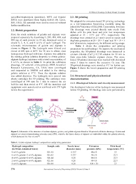
Figure 1. Schematic of the structure of sodium alginate, gelatin, and gelatin-alginate blend in 3D-printed cell-laden dressings. Gelatin and
alginate are semi-interpenetrating networks (semi-IPN), whereby the linear chains of alginate are embedded within the gelatin network,
which decreases the free volume .
[39]
Table 1. Gelatin-alginate hydrogel compositions and printing parameters
Hydrogel composition Sample code Nozzle inner Extrusion Print head
(in 10 mL deionized water) diameter (mm) pressure (kPa) temperature (°C)
8 w/v% gelatin G8-A0 0.337 40±10 22
6 w/v% gelatin+2 w/v% alginate G6-A2 0.337 50±10 22.5
4 w/v% gelatin+4 w/v% alginate G4-A4 0.26 80±10 22.5
2 w/v% gelatin+6 w/v% alginate G2-A6 0.26 80±10 22.5
8 w/v% alginate G0-A8 0.26 100 ± 20 23
276 International Journal of Bioprinting (2022)–Volume 8, Issue 4