Page 285 - IJB-8-4
P. 285
Fayyazbakhsh, et al.


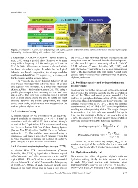
           Figure 2. Fabrication of 3D-printed wound dressings with alginate, gelatin, and human dermal fibroblasts for partial-thickness burn wounds
           followed by 10 min crosslinking with calcium chloride solution.

           a Rheometer (HAAKE™ MARS™, Thermo Scientific,       the crystal. A fresh reference spectrum was recorded after
           MA, USA) using a parallel  plate  (diameter  = 35  mm)   every five scans and deducted from the obtained spectra.
           setup with a frequency of 1 Hz and a gap of 1 mm at   All the recorded spectra were analyzed with OMNIC
           22°C for 300 s. All measurements were performed within   9.2.41  software  (Thermo  Scientific,  MA,  USA),  and
           the  linear viscoelastic  region. To relate  the  rheological   data showed as Transmittance (%) against wavenumber
                                                                  −1
           behavior  and bioink  composition, the storage  modulus   (cm ). The IR Spectrum data from Sigma Aldrich were
           and loss modulus (G’ and G”, respectively) were analyzed   used to identify characteristic chemical bonds in gelatin,
           for the various gelatin: alginate ratios.           alginate, and water.
               The viscosity and shear thinning behavior of the
           non-printed  hydrogels  with  different  ratios  of  gelatin   2.5. Swelling capacity and biodegradation rate
           and alginate  were measured  by a rotational  rheometer   measurement
           (Kinexus, Ultra+, Malvern Instruments Ltd., UK) using a   To determine the further interactions between the wound
           parallel plate setup for shear rate ramp test with a 0.5 mm   and dressing, the swelling capacity and the degradation
           gap at 22°C. The tests were conducted using a solvent   rate  of the 3D-printed  dressings were recorded after
           trap to avoid drying during the test. To relate the shear   soaking  in  phosphate-buffered  saline  (PBS).  Samples
           thinning behavior and bioink composition,  the shear   were dried at room temperature, and the dry weight of the
           stress, shear strain, and shear rate were measured for the   samples was recorded as W  (n = 3). Then, the samples
                                                                                      D
           various gelatin: alginate ratios.                   were immersed in 3 mL PBS at 32°C to reach equilibrium
                                                               swelling and subsequent degradation. The weight changes
           2.4.2. Mechanical testing                           in determined time intervals were recorded for up to
           A uniaxial tensile test was performed on the dog-bone-  7 days as the dressings will stay on the wound for up to
           shaped  scaffolds  of  dimensions  25  ×  50  ×  1  mm  in   7 days. The dressing’s swelling capacity and degradation
                                                       3
           accordance with the ASTM F2150-8 standard. Young’s   rate were calculated using the following equations:
           moduli of the 3D-printed scaffolds were measured using                         W   − W
           the Universal Instron 5969 Dual Column Testing System      Swellingcapacity % () =  max  D  ×100  (1)
                                                                                             W
           (Instron, MA, USA) and the BlueHill Universal Software                             D
           system (n  =  3).  The  scaffolds  were  assessed  using  a                        W −  W Week1
                                                                                                D
           mechanical load frame at a speed of 5 mm/min typical for      Degradationratemgmin(  /  ) =  72460    (2)
                                                                                                    ×
                                                                                                ×
           polymer specimens to measure the modulus of elasticity,
           yield strength, and yield strain of the scaffolds.      where W  is the  initial  dry weight,  W max  is the
                                                                           D
                                                               maximum weight of the scaffolds after immersion, and
           2.4.3. Fourier-transform infrared spectroscopy (FTIR)   W week1  is the weight after 1 week of immersion in PBS.
           A Nicolet  iS50 FTIR spectrophotometer  (Thermo     2.6. Hydration activity test
           Scientific, MA, USA) equipped with a diamond crystal
           cell of attenuated total reflection accessory in the mid-IR   To  measure  the  effect  of  gelatin:  alginate  ratio  on
           region (4000 – 400 cm ). All the spectra were recorded   hydration  activity,  firstly,  the  total  amount  of  water
                              −1
           at a resolution of 4 cm  with 32 scans with a data spacing   in  each  3D-printed  scaffold  was  measured  using
                             −1
           of  0.482 cm . 500 μL of each hydrogel sample were   thermogravimetric analysis on 250°C for 10 min (SDT
                     −1
           loaded on the beam splitter area to make a thin film on   Q600 V20.9 Build 20, Universal V4.5A TA Instruments,
                                       International Journal of Bioprinting (2022)–Volume 8, Issue 4       277

