
Page 11 - IJB-9-1
P. 11
International Journal of Bioprinting Biocompatible materials and Multi Jet Fusion
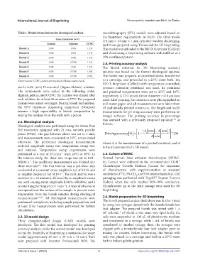
Table 1. Bioink formulations for rheological analysis stereolithography (STL) models were adjusted based on
the bioprinter requirements in Slic3r. The third model
Concentrations (w/v) (10 mm × 10 mm × 1 mm cylinder) was less challenging,
Gelatin Alginate CCNC and it was prepared using Thinkercad for 3D bioprinting.
Bioink A 3.0% 0.5% 1.4% This model was uploaded to the BIO X bioprinter (Cellink)
Bioink B 4.0% 0.5% 1.4% and sliced using a bioprinting software with infill set at a
Bioink C 5.0% 0.5% 1.4% 35% rectilinear pattern.
Bioink D 4.0% 0.75% 1.4% 2.4. Printing accuracy analysis
Bioink E 4.0% 1.0% 1.4% The bioink selection for 3D bioprinting accuracy
Bioink F 4.0% 0.75% 1.0% analysis was based on the former rheological analysis.
Bioink G 4.0% 0.75% 2.0% The bioink was prepared as described above, transferred
Abbreviation: CCNC, carboxymethylated cellulose nanocrystal. to a cartridge, and precooled in a 25°C water bath. The
BIO X bioprinter (Cellink) with temperature-controlled,
sterile 4.6% (w/v) D-mannitol (Sigma-Aldrich) solution. pressure extrusion printhead was used. Its printhead
The components were added in the following order: and printbed temperatures were set to 25°C and 10°C,
alginate, gelatin, and CCNC; the mixture was shaken after respectively. A 22 G needle (inner diameter = 410 µm) was
each addition for at least 30 minutes at 37°C. The prepared used. After printing, the constructs were photographed on
bioinks were mixed overnight. During bioink formulation, millimeter paper, and all measurements were taken from
the EFD Optimum dispensing equipment (Nordson) 15 individually printed constructs. The length and width
ensures a high repeatability of bioink composition by measurements for printing accuracy were performed on
wiping the residues from the walls with a piston. ImageJ software. The printing accuracy in percentage
was assessed with a previously proposed equation as
[38]
2.2. Rheological analysis follows:
Rheological analysis was performed using the Anton Paar
i [
302 rheometer, equipped with 25 mm, smooth, parallel | Amm] − [
A mm | ]
plates (PP25). The gap between plates was set to 1 mm, Printing accuracy % [] = 1 − Amm] * 100
[
and measurements were conducted at 23°C, unless stated
otherwise. The performed rheological measurements where A is the measurement of a printed construct, and A
i
included amplitude sweep test, temperature sweep test, is the measurement of a 3D model.
and rotation. Temperature sweep experiments were
performed at a rate of 2°C·min from 20°C to 40°C. In 2.5. Culture of NHAC
−1
the rotation study, the shear rate range was set to 0.01– Normal human knee articular chondrocytes (NHAC-
200.00 s . The oscillatory measurement was divided into kn, Lonza) were cultured in the recommended CGM™
−1
three intervals . The first interval was a pre-shear step Chondrocyte Growth Medium (Lonza) for expansion
[36]
conducted at a constant strain amplitude (γ) of 0.01% and of chondrocytes, with supplementation at standard
an angular frequency (ω) of 10 s . The next interval was a conditions (37°C, 5% CO , and 95% relative humidity). Cell
−1
2
rest time (t = 10 minutes), followed by an amplitude sweep passaging was performed with TrypLE™ Express Enzyme
test with varying strain amplitude (0.01%–500.00%) and a (Gibco) when the cells reached 80%–90% confluence.
constant angular frequency (1 rad·s ). A layer of silicone oil Chondrocytes up to the sixth passage were used for 3D
−1
was spread over the surface of the sample to prevent water bioprinting.
evaporation from the bioink samples during rheological
measurements [36,37] . All rheological measurements were 2.6. Bioink preparation for 3D bioprinting
performed in triplicate, including sample preparation, and The bioink prepared as described above was further mixed
at least three measurements were performed for further by using two syringes clipped with the female/female luer
calculations. lock adapter. The prepared bioink was mixed with 1 ×
10 cells·mL of bioink in the same way. Specifically, the
7
−1
2.3. 3D model design cells were suspended in 100 µL of chondrocyte medium
Three computer-aided design (CAD) models were and transferred to a syringe, while 1 mL of bioink was
developed. The first model was developed for printing transferred to another syringe; then, the syringes were
accuracy analysis, while the second model was developed clipped with a female/female luer lock adapter prior to
to test the feasibility of bioprinting a meniscus-like shape mixing the content. Before bioprinting, the bioink with
model (approximately 29 mm × 39 mm × 11 mm). Both cells was placed in a cartridge and held in a 25°C water
were prepared with Inventor Professional 2020. The bath to induce gelatin gelation.
Volume 9 Issue 1 (2023) 3 https://doi.org/10.18063/ijb.v9i1.621