
Page 15 - IJB-9-1
P. 15
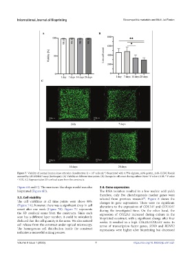
International Journal of Bioprinting                             Biocompatible materials and Multi Jet Fusion


            Figure 7. Viability of normal human knee articular chondrocytes (1 × 10  cells∙mL ) bioprinted with 0.75% alginate_4.0% gelatin_1.4% CCNC bioink
                                                                 −1
                                                           7
            assessed by LIVE/DEAD assay (Invitrogen). (A) Viability at different time points. (B) Changes in cell count during culture. Note: *P value ≤ 0.05; **P value
            ≤ 0.02. (C) Representative 3D confocal scans from the constructs.
            Figure 6A and C). The meniscus-like shape model was also   3.4. Gene expression
            bioprinted (Figure 6D).                            The RNA  isolation  resulted in a  low nucleic  acid yield;
                                                               therefore, only five chondrogenesis marker genes were
            3.3. Cell viability                                selected  from  previous  research .  Figure  8  shows  the
                                                                                          [5]
            The cell viabilities at all time points were above 98%   changes in gene expressions. There were no significant
            (Figure 7A); however, there was a significant drop in cell   alterations to the expressions of COL1A1 and COL10A1
            count after one week (Figure 7B).  Figure 7C represents   during the investigated time. On the other hand, the
            the  3D  confocal  scans  from  the  constructs. Since  each   expression of  COL2A1 increased during culture in the
            scan has a different layer number, it could be mistakenly   bioprinted construct, with a significant change after four
            deduced that the cell quantity is the same. We also noticed   weeks. It resulted in a high  COL2A1/COL1A1 ratio. In
            cell release from the construct under optical microscopy.   terms  of transcription factor  genes,  SOX9  and  RUNX2
            The  homogenous cell  distribution  inside the construct   expressions were higher after bioprinting but decreased
            indicates a successful mixing process.


            Volume 9 Issue 1 (2023)                         7                       https://doi.org/10.18063/ijb.v9i1.621