
Page 144 - IJB-9-1
P. 144
International Journal of Bioprinting BNC-reinforced GelMa enhances property of bioprinted cartilage
3.2.2. SEM examination 0.4 – 0.8 bar pressure were used as printing parameters for
Both hydrogels in BNC/GelMA group and GelMA group subsequent experiments.
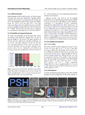
had loose pore structures (Figure 2D). Through further Different models were used to test the printing
analysis of the pore size, it was found that the average pore formability of the composite hydrogel. The presence of BNC
size of the hydrogels in BNC/GelMA group and GelMA led to the extrusion of a stable filament, which, therefore,
group was 172.8 ± 54.19 μm and 126.0 ± 35.21 μm, contributes to a homogenous diameter distribution
respectively (Figure 2E). The scaffold’s pore size of the BNC alongside the length (Figure 4A). When stacked up to 18
group was significantly larger than that of the GelMA group layers, the grid structure formed by the hydrogel could still
(P < 0.0001). The loose pore structure may be conducive be seen (Figure 4B). Then, we printed 0.3 times the size of
to the proliferation and migration of cells in the scaffold . the human jaw model (3.1 × 2.2 × 0.896 cm) (Figure 4C)
[38]
and 0.4 times the size of the human nose model (2.4 × 1.6 ×
3.3. Printability of composite hydrogel 0.576 cm) (Figure 4D). The addition of BNC helped obtain
We tested the printability of the hydrogel with nozzles uniform lines, complete printing structure, clear surface
of different inner diameters ranging from 150 μm to outline, and high fidelity; these characteristics are essential
600 μm (Figure 3). The nozzles with inner diameters of for the construction of scaffolds with precise morphology.
150 μm–210 μm could not be extruded completely, and 3.4. Cell viability and migration
the nozzles with inner diameters of 250 μm–300 μm
were occasionally blocked. When a 400 μm nozzle was 3.4.1. Cell viability
used, the hydrogel could be smoothly extruded under The Calcein AM/PI double staining kit was used to stain
pressures above 0.4 bar. Taking the cell viability and the live and dead cells on the 1 , 4 , and 7 days after
th
th
st
printing accuracy into consideration, a 400 μm nozzle and printing to evaluate the cell viability. Most of the cells in
the scaffolds were dyed green fluorescence, and only a few
cells showed red fluorescence, suggesting high cell viability
in the hydrogels (Figure 5A). ImageJ software was used
for quantitative analysis of the number of live and dead
cells. The percentages of cell viability of the BNC/GelMA
th
th
group on the 1 , 4 , and 7 days were 96.81 ± 1.541%,
st
96.12 ± 0.6627%, and 97.34 ± 1.450%, respectively
(Figure 5B). There was no significant difference in terms
of cell viability between the BNC/GelMA group and the
GelMA group (P > 0.05).
Figure 3. Extrusion test of the bacterial nanocellulose (BNC)/gelatin
methacryloyl (GelMA) hydrogel at 21°C with various pressures and 3.4.2. Cell migration
nozzles. Black indicates that BNC/GelMA cannot be extruded, red
indicates that BNC/GelMA cannot be extruded smoothly, and green To explore whether larger internal pores of the BNC/GelMA
indicates that BNC/GelMA can be extruded smoothly. hydrogel affected the migration of cells in the scaffold, we
A B
C D
Figure 4. Printability test of bacterial nanocellulose (BNC)/gelatin methacryloyl (GelMA) hydrogel. (A) PSH characters printed with BNC/GelMA
hydrogel. (B) Cuboid structure at different layers printed with BNC/GelMA hydrogel. (C) Human mandibular model printed with BNC/GelMA hydrogel.
(D) Human nose model printed with BNC/GelMA hydrogel.
Volume 9 Issue 1 (2023) 136 https://doi.org/10.18063/ijb.v9i1.631