
Page 147 - IJB-9-1
P. 147
International Journal of Bioprinting BNC-reinforced GelMa enhances property of bioprinted cartilage
th
th
th
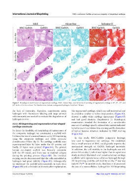
Figure 7. Histological examination of regenerated cartilage. H&E, Alcian blue, and Safranine-O staining of regenerated cartilage at 4 , 8 , 12 , and
th
24 weeks of in vivo culture. The black arrows indicate undegraded hydrogels. Scale bar: 100 μm.
the lack of immunity. Therefore, experiments using The regenerated cartilage, which was well maintained and
hydrogels with fluorescent labeling and large animals in condition similar to before implantation (Figure 8E),
with immunity are needed to evaluate the degradation of showed a milky white cartilage appearance (Figure 8F)
hydrogels in vivo. and had good elasticity (Supplement 2). Histological
examination revealed the formation of a considerable
3.5.3. 3D bioprinting and regeneration of ear-shaped amount of cartilage-specific extracellular matrix indicated
cartilage constructs
by Safranine-O and Alcian blue staining and the formation
To detect the feasibility of morphological maintenance of of typical lacunae structure indicated by H&E staining
the composite hydrogel, we constructed a scaffold with (Figure 8G).
0.4 times the size of a normal human ear by 3D bioprinting
using the composite hydrogel and rabbit auricular In this study, BNC/GelMA composite hydrogel
chondrocytes (Video clip 1). The cartilage scaffolds were was used to regenerate ear cartilage. We confirmed
superimposed layer by layer under the 3D printer, and that a small amount of BNC could greatly improve the
finally, 22 layers were printed (Figure 8A). The printed mechanical strength of GelMA hydrogel materials.
human ear-shaped scaffold was basically consistent In addition, the cell viability in the hydrogels was still
with the 3D model, and there was no material collapse above 95% on day 7, which was higher than in a previous
during the printing process (Figure 8B). The live/dead study, in which Markstedt et al. constructed ear cartilage
staining results demonstrated that the cells embedded in scaffolds with alginate/nano-cellulose hydrogel through
th
hydrogels had good viability (Figure 8C). Subsequently, 3D bioprinting, and the cell viability on the 7 day was
we implanted the scaffold subcutaneously in nude mice only 85.7% [44] . In another study, Martínez et al. proposed
(Figure 8D) and collected it at 24 weeks after implantation. the use of a nanocellulose hydrogel for 3D bioprinting
Volume 9 Issue 1 (2023) 139 https://doi.org/10.18063/ijb.v9i1.631