
Page 145 - IJB-9-1
P. 145
International Journal of Bioprinting BNC-reinforced GelMa enhances property of bioprinted cartilage
A
B D
C
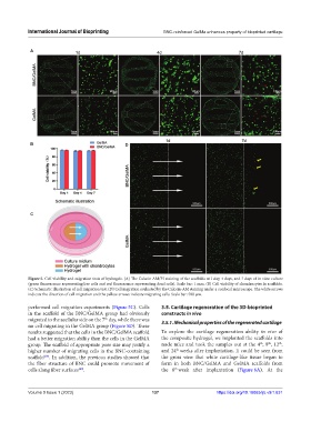
Figure 5. Cell viability and migration tests of hydrogels. (A) The Calcein AM/PI staining of the scaffolds at 1 day, 4 days, and 7 days of in vitro culture
(green fluorescence representing live cells and red fluorescence representing dead cells). Scale bar: 1 mm. (B) Cell viability of chondrocytes in scaffolds.
(C) Schematic illustration of cell migration test. (D) Cell migration evaluated by the Calcein-AM staining under a confocal microscope. The white arrows
indicate the direction of cell migration and the yellow arrows indicate migrating cells. Scale bar: 500 μm.
performed cell migration experiments (Figure 5C). Cells 3.5. Cartilage regeneration of the 3D-bioprinted
in the scaffold of the BNC/GelMA group had obviously constructs in vivo
migrated to the acellular side on the 7 day, while there was 3.5.1. Mechanical properties of the regenerated cartilage
th
no cell migrating in the GelMA group (Figure 5D). These
results suggested that the cells in the BNC/GelMA scaffold To explore the cartilage regeneration ability in vivo of
had a better migration ability than the cells in the GelMA the composite hydrogel, we implanted the scaffolds into
th
group. The scaffold of appropriate pore size may justify a nude mice and took the samples out at the 4 , 8 , 12 ,
th
th
th
higher number of migrating cells in the BNC-containing and 24 weeks after implantation. It could be seen from
scaffold . In addition, the previous studies showed that the gross view that white cartilage-like tissue began to
[39]
the fiber structure of BNC could promote movement of form in both BNC/GelMA and GelMA scaffolds from
cells along fiber surfaces . the 8 week after implantation (Figure 6A). At the
[40]
th
Volume 9 Issue 1 (2023) 137 https://doi.org/10.18063/ijb.v9i1.631