
Page 141 - IJB-9-1
P. 141
International Journal of Bioprinting BNC-reinforced GelMa enhances property of bioprinted cartilage
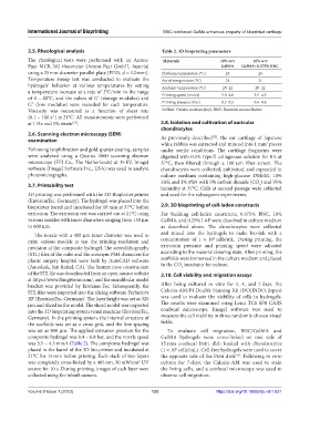
2.5. Rheological analysis Table 2. 3D bioprinting parameters
The rheological tests were performed with an Anton- Materials 10% w/v 10% w/v
Paar MCR 302 rheometer (Anton-Paar GmbH, Austria) GelMA GelMA+0.375% BNC
using a 25 mm diameter parallel plate (PP25, d = 1.0 mm). Platform temperature (°C) 25 25
Temperature sweep test was conducted to evaluate the Barrel temperature (°C) 21 21
hydrogels’ behavior at various temperatures by setting Ambient temperature (°C) 20–22 20–22
a temperature increase at a rate of 2°C/min in the range
of 0 – 40°C, and the values of G’ (storage modulus) and Printing speed (mm/s) 5.0–8.0 3.5–4.5
G” (loss modulus) were recorded for each temperature. Printing pressure (Bar) 0.2–0.5 0.4–0.8
Viscosity was measured as a function of shear rate GelMA: Gelatin methacryloyl, BNC: Bacterial nanocellulose
(0.1 – 100 s ) at 21°C. All measurements were performed
-1
at 1 Hz and 1% strain . 2.8. Isolation and cultivation of auricular
[31]
chondrocytes
2.6. Scanning electron microscopy (SEM) [32]
examination As previously described , the ear cartilage of Japanese
white rabbits was extracted and minced into 1 mm pieces
3
Following lyophilization and gold sputter coating, samples under sterile conditions. The cartilage fragments were
were analyzed using a Quanta 2000 scanning electron digested with 0.2% type II collagenase solution for 8 h at
microscope (FEI Co., The Netherlands) at 15 kV. ImageJ 37°C, then filtered through a 100 μm filter screen. The
software (ImageJ Software Inc., USA) was used to analyze chondrocytes were collected, cultivated, and expanded in
photomicrographs. culture medium containing high-glucose DMEM, 10%
FBS, and 1% PSN with 5% carbon dioxide (CO ) and 95%
2.7. Printability test humidity at 37°C. Cells at second passage were collected
2
3D printing was performed with the 3D-Bioplotter printer and used for the subsequent experiments.
(EnvisionTec, Germany). The hydrogel was placed into the
bioprinter barrel and incubated for 10 min at 37°C before 2.9. 3D bioprinting of cell-laden constructs
extrusion. The extrusion test was carried out at 21°C using For building cell-laden constructs, 0.375% BNC, 10%
various nozzles with inner diameters ranging from 150 μm GelMA, and 0.25% LAP were dissolved in culture medium
to 600 μm. as described above. The chondrocytes were collected
The nozzle with a 400 μm inner diameter was used to and mixed into the hydrogels to make bio-ink with a
7
print various models to test the printing resolution and concentration of 1 × 10 cells/mL. During printing, the
precision of the composite hydrogel. The stereolithography extrusion pressure and printing speed were adjusted
(STL) files of the cube and the acronym PSH characters for according to the material drawing state. After printing, the
plastic surgery hospital were built by AutoCAD software scaffolds were immersed in the culture medium and placed
(Autodesk, San Rafael, CA). The human nose construction in the CO incubator for culture.
2
of the STL file was downloaded from an open-source website 2.10. Cell viability and migration assays
at https://www.thingiverse.com/, and the mandibular model
bracket was provided by Envision-Tec. Subsequently, the After being cultured in vitro for 1, 4, and 7 days, the
STL files were imported into the slicing software Perfactory Calcein-AM/PI Double Staining Kit (DOJINDO, Japan)
RP (EnvisionTec, Germany). The layer height was set as 320 was used to evaluate the viability of cells in hydrogels.
μm and sliced in the model. The sliced model was imported The results were examined using Leica TCS SP8 CARS
into the 3D bioprinting system visual machine (EnvisionTec, confocal microscope. ImageJ software was used to
Germany). In the printing system, the internal structure of measure the cell viability in three randomly chosen visual
the scaffolds was set as a cross grid, and the line spacing fields.
was set as 800 μm. The applied extrusion pressure for the To evaluate cell migration, BNC/GelMA and
composite hydrogel was 0.4 – 0.8 bar, and the nozzle speed GelMA hydrogels were cross-linked on one side of
was 3.5 – 4.5 mm/s (Table 2). The composite hydrogel was 15 mm confocal Petri dish loaded with chondrocytes
placed in the barrel of the 3D bio-printer and incubated at (1 × 10 cells/mL). Cell-free hydrogels were used to cover
6
21°C for 15 min before printing. Each stack of two layers the opposite side of the Petri dish [33] . Following in vitro
was completely cross-linked by a 405 nm, 30 mW/cm UV culture for 7 days, the Calcein-AM was used to stain
2
source for 10 s. During printing, images of each layer were the living cells, and a confocal microscope was used to
collected using the inbuilt camera. observe cell migration.
Volume 9 Issue 1 (2023) 133 https://doi.org/10.18063/ijb.v9i1.631