
Page 176 - IJB-9-1
P. 176
International Journal of Bioprinting Lumen-forming colorectal cancer organoids
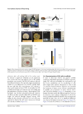
Figure 6. Three-dimensional printed construct using IIFK:IDP hydrogel. (A) In-house bioprinting setup with robotic arm setup. (B) Top and side views
of printed construct of IIFK:IDP (5:1) at 8 mM and 10 mM. (C) Printed constructs at days 1 and 7 of bioprinting of IIFK:IDP (5:1) at 8 mM. (D) Bioprint-
ability assessment by cell proliferation assay in the IIFK:IDP construct after 1 and 7 days of 3D bioprinting and culture.
adherence after cell seeding (44% of the surface area) 3.4. Characterization of CRC cells in scaffolds
but showed a significant reduction of cell population In order to determine whether our peptide scaffolds
after mechanical stress was applied (57% of the seeded contribute to the initial organization of cells into organoids,
population was retained). The IIFK:IDP mixtures showed single CRC cells were embedded into each peptide, and
two different behaviors. The one with a high concentration the F-actin cytoskeleton and the morphology of cells
of IDP (1:1) resembled that of the IKVAV scaffold, while were imaged with confocal fluorescence at days 4 and
the one with high IIFK concentration (10:1) showed a 7 to monitor lumen formation. Moreover, we evaluated
closer performance to that of IIFK. Incidentally, the IDP the formation of lumen across different concentrations
scaffold showed high cell retention after cell seeding and of peptides and compared it to lumen formed within
the application of mechanical stress. Taken together, SAP cells cultured in Matrigel (Figure 5). Morphology of the
hydrogels may facilitate physical/mechanical retention luminal structures across the different peptides varied as
of cells in the fiber network, while IDP and IKVAV may we observed a more ordered configuration of cells in the
trigger cell adhesion through integrin binding. SEM biofunctionalized and IKVAV peptides. The concentrations
images of cells cultured in IIFK:IDP (1:1) at 8 mM were used did not play a key role as no significant difference
captured to observe the cell–matrix interactions formed was observed in lumen formation (Figure 5A, Figure S5).
after one day of culture (Figure 4C). Figure 5B shows the difference in the diameter of lumen
V
Volume 9 Issue 1 (2023)olume 9 Issue 1 (2023) 168 https://doi.org/10.18063/ijb.v9i1.633