
Page 175 - IJB-9-1
P. 175
International Journal of Bioprinting Lumen-forming colorectal cancer organoids
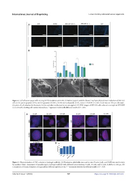
Figure 4. Cell adhesion assays with varying SAP formulations at 8 mM. (A) Before (upper) and after (lower) mechanical detachment induction of SW1222
cells on the parent peptide (IIFK), laminin peptide (IKVAV), IKVAV-derived peptide (IDP), and an IIFK:IDP (1:1) Mix. Scale bars are 100 μm. (B) Quan-
tification of cells attached to the matrix before and after mechanical stress was applied. (C) SEM images of SW1222 cells cultured overnight in IIFK:IDP
(1:1) at 8 mM, showing cell–matrix interactions. * represents statistically different results at P < 0.05.
Figure 5. Characterization of CRC colonies in hydrogel scaffolds. (A) Rhodamine phalloidin was used to stain F-actin (red), and DAPI was used to stain
the nucleus (blue). Assessment of morphology in hydrogel scaffolds with different concentrations: 8 mM, 10 mM, and 12 mM. Scale bar is 100 μm. (B)
Comparison of lumen diameters of organoids in different peptides at day 7. * represents statistically different results at P < 0.05.
Volume 9 Issue 1 (2023)olume 9 Issue 1 (2023)
V 167 https://doi.org/10.18063/ijb.v9i1.633