
Page 174 - IJB-9-1
P. 174
International Journal of Bioprinting Lumen-forming colorectal cancer organoids
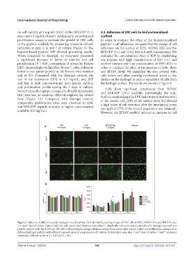
on cell viability at 3 mg/mL (CGC of the IIFK:IDP (1:1) 3.3. Adhesion of CRC cells to biofunctionalized
Mix) and 4.5 mg/mL (8 mM). Additionally, we performed scaffold
proliferation assays to estimate the growth of CRC cells In order to evaluate the effect of the biofunctionalized
in the peptide scaffolds by measuring resazurin cellular peptide in cell adherence, we quantified the change of cell
reduction at days 1, 4, and 7 of culture (Figure 3). The adherence on the surface of IIFK, IKVAV, IDP, and the
laminin-based peptide IDP showed promising results. IIFK:IDP (1:1) and (10:1) Mix at 8 mM concentration. We
When compared to Matrigel, no treatment presented evaluated the concentration effect of IDP by establishing
a significant decrease in terms of viability and cell one mixture with high concentration of IDP (1:1) and
proliferation (P < 0.01, comparison of means by Fishers another mixture with low concentration of IDP (10:1) in
LSD). Interestingly, we find that, by day 7, cells proliferate order to compare the effect of the presence of IIFK fibers
better in our parent peptide at the lowest concentration and IKVAV motif. We quantified the area covered with
and in 2D. Compared with the Matrigel control, the cells before and after exerting mechanical stress in the
rest of the treatments (IIFK at 4.5 mg/mL, and IDP surface on the hydrogel to induce separation of cells from
and Mix at both concentrations) have similar viability the hydrogel surface. The results are shown in Figure 4.
and proliferation profile during the 7 days of culture. Cells show significant detachment from IKVAV
Nevertheless, the high percentage of cell viability indicates and IIFK:IDP (10:1) scaffolds. Interestingly, the non-
that there was no cytotoxic effect throughout the culture biofunctionalized peptide IIFK had a lower initial retention
time (Figure 3A). Compared with Matrigel control, of the seeded cells (18% of the surface area) but showed
comparable proliferation rates were observed in IIFK a high index of cell retention after the mechanical stress
and IIFK:IDP peptide mixtures at higher concentration was applied (77% of the seeded population was retained).
scaffolds (4.5 mg/mL). However, the IKVAV scaffold induced an increase in cell
Figure 3. Influence of different peptide hydrogels on cell viability. (A) Cell viability staining images of CRC cells in IIFK, IKVAV, IDP, and IIFK:IDP after
1, 4, and 7 days of culture. Calcein AM (live cells, green) and ethidium homodimer-1 (dead cells, red) were used to stain the cells. Matrigel was used as a
positive control. Scale bar is 100 μm. (B) (left) Cell viability percentages obtained using a fluorescence plate reader; (right) cell proliferation assessment in
different hydrogel scaffolds with different concentrations in comparison to 2D culture. Proliferation assay after 1 and 7 days of culture. * and ** represent
statistically different results at P < 0.05 and P < 0.01.
Volume 9 Issue 1 (2023)olume 9 Issue 1 (2023)
V 166 https://doi.org/10.18063/ijb.v9i1.633