
Page 184 - IJB-9-1
P. 184
International Journal of Bioprinting PEEK skull implant in cranioplasty
machine (printer model: Surgeon Pro) automatically Table 1. Comparison of mechanical properties between the
printed the PEEK skull implant layer-by-layer, as shown skull and 3D-printed PEEK implants
in Figure 2C. For the fabrication of the as-designed PEEK Mechanical properties Skull [13] 3D-printed PEEK implant
skull implant, the printing parameters of FFF such as layer
thickness, nozzle diameter, bed and chamber temperature, Elastic modulus (GPa) 8.51 3.45
nozzle temperature, and printing speed were set at Tensile strength (MPa) 67.73 96
0.2 mm, 0.4 mm, 20°C, 430°C, and 40 mm/s, respectively. Flexural strengths (MPa) 82 154
The macroscopic image of the FFF-printed PEEK prosthesis Impact toughness (kJ/m ) 49 80
2
skull is shown in Figure 2D.
The weight of the FFF-printed PEEK skull prosthesis Then, we compared the degree of integration of the
was approximately 42.79 g. The size of the skull implant skull made by stereolithography appearance technology
reached 12 cm × 10 cm. Before clinical application, with the titanium mesh and PEEK implants from various
several mechanical properties were tested by a third-party angles before surgery (Figure 3A–F). We found that
inspection institution (National Additive Manufacturing the newly regenerated bone was tightly bound to the
Product Quality Supervision and Testing Center). implanted titanium mesh, and a clear gap was formed
Subsequently, we further compared the relevant parameters between the skull and the titanium mesh in the temporal
with previous reports on the mechanical properties of area (Figure 3A–C). In order to avoid further damage to
the human skull . The elastic modulus of the skull and the newly regenerated bone on the dura after peeling the
[13]
3D-printed PEEK implants are 8.51 GPa and 3.45 GPa, titanium mesh, we appropriately increased the curvature
the tensile strengths are 67.73 MPa and 96 MPa, and the of the central part of the PEEK implant. Although some
flexural strengths are 82 MPa and 154 MPa (Table 1). The scholars have considered that the increase in curvature
printed PEEK implant was scanned with a 3D scanner may cause the collapse of patient’s scalp incision due to
(XTOM-MATRIX) to obtain the actual size of the implant. excessive tension , this patient had no significant incision
[12]
Then, the deviation between the design model and the complications. In addition, during the production process,
printed PEEK implant was analyzed by the built-in analysis we filled the gaps in the temporal region according to the
software (Figure 2E and F). patient’s skull anatomy (Figure 3B and E). Finally, we found
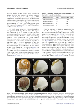
Figure 3. The preoperative matching between skull defects model and PEEK material. (A–C) The skull defects model made by stereolithography
technology shows the positional relationship between the skull and titanium. The newly regenerated bone (indicated by the blue arrow) merged with the
titanium mesh. In the temporal area (B), a clear gap between the titanium mesh and skull (red arrow) can be observed. (D–F) The PEEK implant was
fabricated by FFF technology for preoperative matching. The inner side of the blue line is the PEEK implant of uniform thickness, and the outer side is the
thinning wing (D). In the temporal part (E), the PEEK skull implant shows a tight fit for the skull defects model (red arrow) in the preoperative matching.
V
Volume 9 Issue 1 (2023)olume 9 Issue 1 (2023) 176 https://doi.org/10.18063/ijb.v9i1.634