
Page 185 - IJB-9-1
P. 185
International Journal of Bioprinting PEEK skull implant in cranioplasty
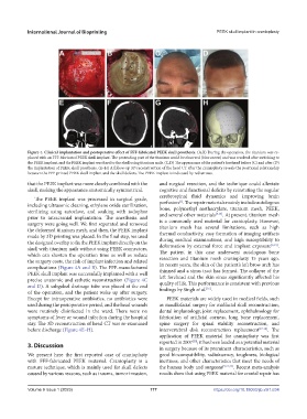
Figure 4. Clinical implantation and postoperative effect of FFF-fabricated PEEK skull prosthesis. (A,B) During the operation, the titanium was re-
placed with an FFF-fabricated PEEK skull implant. The protruding part of the titanium could be observed (blue arrow) and was resolved after switching to
the PEEK implant, and the PEEK implant was fixed to the skull using titanium nails. (C,D) The appearance of the patient’s forehead before (C) and after (D)
the implantation of PEEK skull prosthesis. (E–H) A follow-up 3D reconstruction of the head CT after the cranioplasty reveals the positional relationship
between the FFF-printed PEEK skull implant and the skull defects. The PEEK implant is indicated by red arrows.
that the PEEK implant was more closely combined with the and surgical resection, and the technique could alleviate
skull, making the appearance anatomically symmetrical. cognitive and functional deficits by reinstating the regular
cerebrospinal fluid dynamics and improving brain
The PEEK implant was processed in surgical grade, [1]
including ultrasonic cleaning, ethylene oxide sterilization, perfusion . The repair materials mainly include autologous
bone, polymethyl methacrylate, titanium mesh, PEEK,
sterilizing using autoclave, and soaking with iodophor and several other materials [3,14] . At present, titanium mesh
prior to intracranial implantation. The anesthesia and is a commonly used material for cranioplasty. However,
surgery were going well. We first separated and removed titanium mesh has several limitations, such as high
the deformed titanium mesh, and then, the PEEK implant thermal conductivity, easy formation of imaging artifacts
made by 3D printing was placed. In the final step, we used during medical examinations, and high susceptibility to
the designed overlap to fix the PEEK implant directly on the deformation by external force and implant exposure [3,15] .
skull with titanium nails without using PEEK connectors, The patient in this case underwent autologous bone
which can shorten the operation time as well as reduce resection and titanium mesh cranioplasty 15 years ago.
the surgery costs, the risk of implant infection and related In recent years, the skin of the patient’s left brow arch has
complications (Figure 4A and B). The FFF-manufactured thinned and a sinus tract has formed. The collapse of the
PEEK skull implant was successfully implanted with a well left forehead and the skin sinus significantly affected his
precise anatomic and esthetic reconstruction (Figure 4C quality of life. This performance is consistent with previous
and D). A subgaleal drainage tube was placed at the end findings by Singh et al. .
[16]
of the operation, and the patient woke up after surgery.
Except for intraoperative antibiotics, no antibiotics were PEEK materials are widely used in medical fields, such
used during the perioperative period, and the head wounds as maxillofacial surgery for midfacial skull reconstruction,
were routinely disinfected in the ward. There were no dental implantology, joint replacement, ophthalmology for
symptoms of fever or wound infection during the hospital fabrication of artificial corneas, long bone replacement,
stay. The 3D reconstruction of head CT was re-examined spine surgery for spinal stability reconstruction, and
before discharge (Figure 4E–H). intervertebral disk reconstruction replacement [17-19] . The
application of PEEK material for cranioplasty was first
[20]
3. Discussion reported in 2007 ; it has been lauded as a potential material
in surgery because of its prominent characteristics, such as
We present here the first reported case of cranioplasty good biocompatibility, radiolucency, toughness, biological
with FFF-fabricated PEEK material. Cranioplasty is a inertness, and other characteristics that meet the needs of
mature technique, which is mainly used for skull defects the human body and surgeons [5,21,22] . Recent meta-analysis
caused by various reasons, such as trauma, tumor invasion, results show that using PEEK material for cranial repair has
V 177 https://doi.org/10.18063/ijb.v9i1.634
Volume 9 Issue 1 (2023)olume 9 Issue 1 (2023)