
Page 224 - IJB-9-1
P. 224
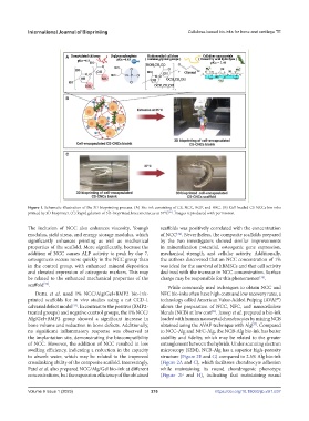
International Journal of Bioprinting                         Cellulose-based bio-inks for bone and cartilage TE


            Figure 1. Schematic illustration of the 3D bioprinting process. (A) Bio-ink consisting of CS, NCC, BGP, and HEC. (B) Cell-loaded CS-NCCs bio-inks
            printed by 3D bioprinter. (C) Rapid gelation of 3D-bioprinted knee meniscus at 37°C . Images reproduced with permission.
                                                                  [31]

            The inclusion of NCC also enhances  viscosity,  Young’s   scaffolds was positively correlated with the concentration
            modulus, yield stress, and energy storage modulus, which   of NCC . Nevertheless, the composite scaffolds prepared
                                                                     [34]
            significantly enhances printing as well as mechanical   by the two investigators showed similar improvements
            properties of the scaffold. More significantly, because the   in mineralization potential, osteogenic gene expression,
            addition of NCC causes ALP activity to peak by day 7,   mechanical  strength,  and  cellular  activity.  Additionally,
            osteogenesis occurs more quickly in the NCC group than   the authors discovered that an NCC concentration of 1%
            in the control group, with enhanced mineral deposition   was ideal for the survival of hBMSCs and that cell activity
            and elevated expression of osteogenic markers. This may   declined with the increase in NCC concentration. Surface
            be related to the enhanced mechanical properties of the   charge may be responsible for this phenomenon .
                                                                                                     [35]
            scaffold .
                  [32]
                                                                  While commonly used techniques to obtain NCC and
               Dutta  et  al. used 1% NCC/Alg/Gel+BMP2 bio-ink-  NFC bio-inks often have high costs and low recovery rates, a
            printed  scaffolds  for  in vivo  studies  using  a  rat  CCD-1   technology called American Value-Added Pulping (AVAP®)
            calvarial defect model . In contrast to the positive (BMP2-  allows the preparation of NCC, NFC, and nanocellulose
                             [33]
            treated groups) and negative control groups, the 1% NCC/  blends (NCB) at low cost . Jessop et al. prepared a bio-ink
                                                                                  [36]
            Alg/Gel+BMP2 group showed a significant increase in   loaded with human nasoseptal chondrocytes by mixing NCB
            bone volume and reduction in bone defects. Additionally,   obtained using the AVAP technique with Alg . Compared
                                                                                                  [37]
            no significant inflammatory response was observed at   to NCC-Alg and NFC-Alg, the NCB-Alg bio-ink has better
            the implantation site, demonstrating the biocompatibility   stability  and  fidelity,  which  may  be  related  to  the  greater
            of NCC. However, the addition of NCC resulted in low   entanglement between the hybrids. Under scanning electron
            swelling efficiency, indicating a reduction in the capacity   microscopy (SEM), NCB-Alg has a superior high-porosity
            to absorb water, which may be related to the improved   structure (Figure 2E and G) compared to 2.5% Alg bio-ink
            crosslinking ability of the composite scaffold. Interestingly,   (Figure 2A and C), which facilitates chondrocyte adhesion
            Patel et al. also prepared NCC/Alg/Gel bio-ink at different   while maintaining its round chondrogenic phenotype
            concentrations, but the expansion efficiency of the obtained   (Figure 2F and  H), indicating that maintaining round


            Volume 9 Issue 1 (2023)olume 9 Issue 1 (2023)
            V                                              216                      https://doi.org/10.18063/ijb.v9i1.637