
Page 88 - IJB-9-1
P. 88
International Journal of Bioprinting Progress in bioprinting of bone
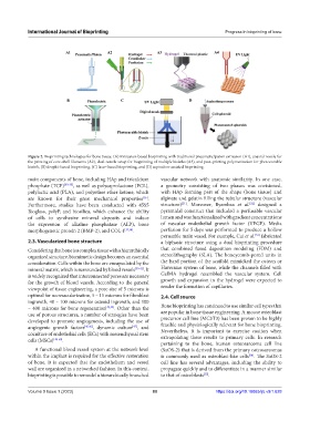
Figure 2. Bioprinting technologies for bone tissue. (A) Extrusion-based bioprinting with traditional pneumatic/piston extrusion (A1), coaxial nozzle for
the printing of core-shell filaments (A2), dual-nozzle setup for bioprinting of multiple bioinks (A3), and post-printing polymerization for photocurable
bioink, (B) droplet-based bioprinting, (C) laser-based bioprinting, and (D) aspiration-assisted bioprinting.
main components of bone, including HAp and tricalcium vascular network with anatomic similarity. In one case,
phosphate (TCP) [33-35] , as well as polycaprolactone (PCL), a geometry consisting of two phases was envisioned,
polylactic acid (PLA), and polyether ether ketone, which with HAp forming part of the shape (bone tissue) and
are known for their great mechanical properties . alginate and gelatin filling the tubular structure (vascular
[36]
[51]
[52]
Furthermore, studies have been conducted with 45S5 structure) . Moreover, Byambaa et al. designed a
Bioglass, polyP, and biosilica, which enhance the ability pyramidal construct that included a perfusable vascular
of cells to synthesize mineral deposits and induce lumen and was functionalized with gradient concentrations
the expression of alkaline phosphatase (ALP), bone of vascular endothelial growth factor (VEGF). Media
morphogenetic protein 2 (BMP-2), and COL-I [37,38] . perfusion for 5 days was performed to produce a hollow
perusable main vessel. For example, Cui et al. fabricated
[53]
2.3. Vascularized bone structure a biphasic structure using a dual bioprinting procedure
Considering that bone is a complex tissue with a hierarchically that combined fused deposition modeling (FDM) and
organized structure, biomimetic design becomes an essential stereolithography (SLA). The honeycomb-pored units in
consideration. Cells within the bone are encapsulated by the the hard portion of the scaffold mimicked the osteon or
mineral matrix, which is surrounded by blood vessels [39-42] . It Haversian system of bone, while the channels filled with
is widely recognized that interconnected pores are necessary GelMA hydrogel resembled the vascular system. Cell
for the growth of blood vessels. According to the general growth and expansion in the hydrogel were expected to
viewpoint of tissue engineering, a pore size of 5 microns is render the formation of capillaries.
optimal for neovascularization, 5 – 15 microns for fibroblast 2.4. Cell source
ingrowth, 40 – 100 microns for osteoid ingrowth, and 100
– 400 microns for bone regeneration [43,44] . Other than the Bone bioprinting has continued to use similar cell types that
use of porous structures, a number of strategies have been are popular in bone tissue engineering. A mouse osteoblast
developed to promote angiogenesis, including the use of precursor cell line (MC3T3) has been proven to be highly
[47]
angiogenic growth factors [45,46] , dynamic culture , and feasible and physiologically relevant for bone bioprinting.
coculture of endothelial cells (ECs) with mesenchymal stem Nevertheless, it is important to exercise caution when
extrapolating these results to primary cells. In research
cells (MSCs) [48-50] .
pertaining to the bone, human osteosarcoma cell line
A functional blood vessel system at the network level (SaOS-2) that is derived from the primary osteosarcomas
within the implant is required for the effective restoration is commonly used as osteoblast-like cells . The SaOS-2
[54]
of bone. It is expected that the endothelium and vessel cell line has several advantages, including the ability to
wall are organized in a networked fashion. In this context, propagate quickly and to differentiate in a manner similar
bioprinting is possible to remodel a hierarchically branched to that of osteoblasts .
[55]
Volume 9 Issue 1 (2023) 80 https://doi.org/10.18063/ijb.v9i1.628