
Page 91 - IJB-9-1
P. 91
International Journal of Bioprinting Progress in bioprinting of bone
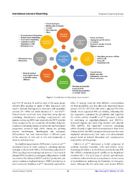
Figure 3. Considerations for bioprinting a bone construct.
and COL-II staining. In another work of the same group, effect of varying materials with different concentrations
different fiber spacings or angles of fiber deposition were on their printability, and thus effectively bioprinted mouse
used to fabricate heterogeneous constructs with adjustable calvaria 3T3-E1 (MC3T3) cells within alginate-PVA-HAp
[81]
porosity (35 – 66%) and elastic modulus (4.7 – 6.6 kPa) . bioink, which supported 96% cell viability, indicating that
Osteochondral constructs were bioprinted using alginate the suspension optimized the printability and improved
containing chondrocytes (cartilage compartment) and the cellular activity. Cunniffe et al. [111] generated a bioink
alginate containing MSCs and osteoinductive BCP particles by combining an arginylglycylaspartic acid (RGD)-γ-
(bone compartment). As a result, the cell viability of alginate irradiated alginate and nano-HAp blended with plasmid
constructs containing human chondrocytes and osteogenic DNA (pDNA). The bioprinted constructs containing
progenitors remained high (90%) during the printing pDNA showed a higher level of mineralization than those
process. Furthermore, chondrogenic and osteogenic without pDNA. The MSC-encapsulated constructs that were
differentiation has been demonstrated in different parts implanted subcutaneously into nude mice demonstrated
of the construct in vitro and in vivo (subcutaneously in greater levels of mineral deposition and vascularization
immune-deficient mice). compared to cell-free groups.
In a study using a pneumatic EBB system, Loozen et al. [110] Cidonio et al. [112] developed a bioink composed of
developed porous or solid constructs containing alginate synthetic nanoclay (Laponite, LPN) and GelMA, which
hydrogel combined with MSCs, BCP particles, and plasmid- showed good fidelity and interlinked porosity during EBB.
DNA-encoding bone morphogenetic protein-2. As a result of The hBMSCs maintained good viability in the bioprinted
plasmid DNA transfection, osteogenic differentiation of cells construct (86 ± 10% at 21 days of culture), and cell-laden
was observed by enhanced BMP-2 and ALP production, and constructs cultured without dexamethasone showed areas
porous constructs displayed superior BMP-2 production to of mineralization, indicating the formation of osteogenic
solid constructs. Bendtsen et al. extensively explored the tissue. Subsequently, the same team used human BMSCs
[83]
Volume 9 Issue 1 (2023) 83 https://doi.org/10.18063/ijb.v9i1.628