
Page 141 - IJB-9-2
P. 141
International Journal of Bioprinting Hybrid biofabrication of neurosecretory structures
the tissue morphology and organ function in vivo over an hydrogel support was unstable, disbanded, and broken,
extended period, all of which are urgent problems that especially when the number of printing layers was low, and
need to be solved in translational medicine research [37,38] . it was difficult to maintain the initial shape (Figure 1A).
The tissue structure of real organs contains not only cells The whole arrangement of the hybrid hydrogel scaffolds
and matrix, but also fibrous connective tissue, which could was observed under an optical microscope, and the print
divide organs into subfunctional units and play a supporting fidelity was high. Moreover, under the cover of nanofibers,
role in maintaining organ morphology . 3D-bioprinted the shape remained good (Figure 1B), and the distributed
[39]
hydrogels can build 3D spatial structures and extracellular neuroendocrine cells in the hydrogel were clearly visible
matrix components for cell survival in vitro, and participate (Figure 1C). As shown by SEM, even after 7 days of culture,
in the construction of tissue-like structures. However, the shape of the hybrid scaffold was well maintained and
hydrogels have poor structural strength and are prone to still covered with fibers (Figure 1D and E). To improve the
quick degradation and losing their original 3D structure. hydrophilicity and biosafety of electrospinning without
Therefore, this method is unable to provide a sustained affecting the strength of the nanofibers, we selected
and stable support for organ-like models in vitro. To better PLLA and gelatin as the raw materials of electrospinning
simulate the organ structure, we introduce electrospun and optimized the ratio and screened the solvents with
PLLA/gelatin nanofiber to generate an organ structure on relatively low residual toxicity. Since most organic solvents
the basis of hybrid biological manufacturing, in which the are toxic, we prefer solvents that are highly volatile and less
nanofibers have a uniform diameter distribution, good toxic to cells . Comparing the effect and residual toxicity
[32]
mechanical properties, hydrophilicity, degradability, and of HFIP, dichloromethane, and acetone electrospinning,
good biocompatibility, without affecting cell activity and it was determined that HFIP had the lowest toxicity
function. Therefore, electrospun PLLA/gelatin nanofiber (Figure S1), and the cytotoxicity of PLLA/gelatin extract
hybrid biological manufacturing realizes the strength and was not significantly different from that of the control
stability of the neurosecretory tissue structure of hybrid group (P < 0.05). By optimizing the ratio, the PLLA/gelatin
biofabrication in vitro. mass fraction ratio of 8:3 was used in electrospinning.
The water contact angles of PLLA/gelatin, PLLA, and
3.1. Hybrid biofabrication of neurosecretory gelatin were 87.75 ± 4.41°, 134.48 ± 3.43°, and 19.43 ±
structures 12.18°, respectively. The results showed that PLLA/gelatin
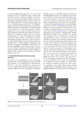
The process of neuroendocrine structure construction has good hydrophilicity and meets the requirements
involved a layer-by-layer “hamburger sandwich” structure of cell adhesion and growth (Figure 2A and B). When
superimposed by 3D bioprinting and electrospinning the nanofiber membrane was immersed in PBS at 50°C,
(Scheme 1). The hybrid biofabrication conferred good the comparison of the relative tensile strength changes
structural stability and could keep the structure intact for a at different times of degradation shows that there is no
long time, while the structure of the simple 3D-bioprinted significant difference in the stress-strain tensile strength of
A B C
F
D E
Scheme 1. Combining 3D bioprinting and electrospinning for hybrid biofabrication.
Volume 9 Issue 2 (2023) 133 https://doi.org/10.18063/ijb.v9i2.659